


Optimising the Use of Contemporary Invasive Imaging for PCI
Published: 06 July 2021
-
Views:
 28982
28982
-
Likes:
 7
7
-
Views:
28982
-
Likes:
7
-
 Up Next
-
 18m 47sPart 1 | Session 1 Welcome and The Science Behind the Pictures Joost Daemen, Margaret McEntegart, Paolo Canova
-
 20mPart 1 | Session 2 How Intravascular Imaging will Change your PCI Strategy Joost Daemen, Margaret McEntegart, Joost Daemen
-
 14m 58sPart 1 | Session 3 Post PCI Optimization Joost Daemen, Margaret McEntegart, Paolo Canova
-
 22m 52sPart 1 | Session 4 Faculty Discussion Joost Daemen, Paolo Canova
-
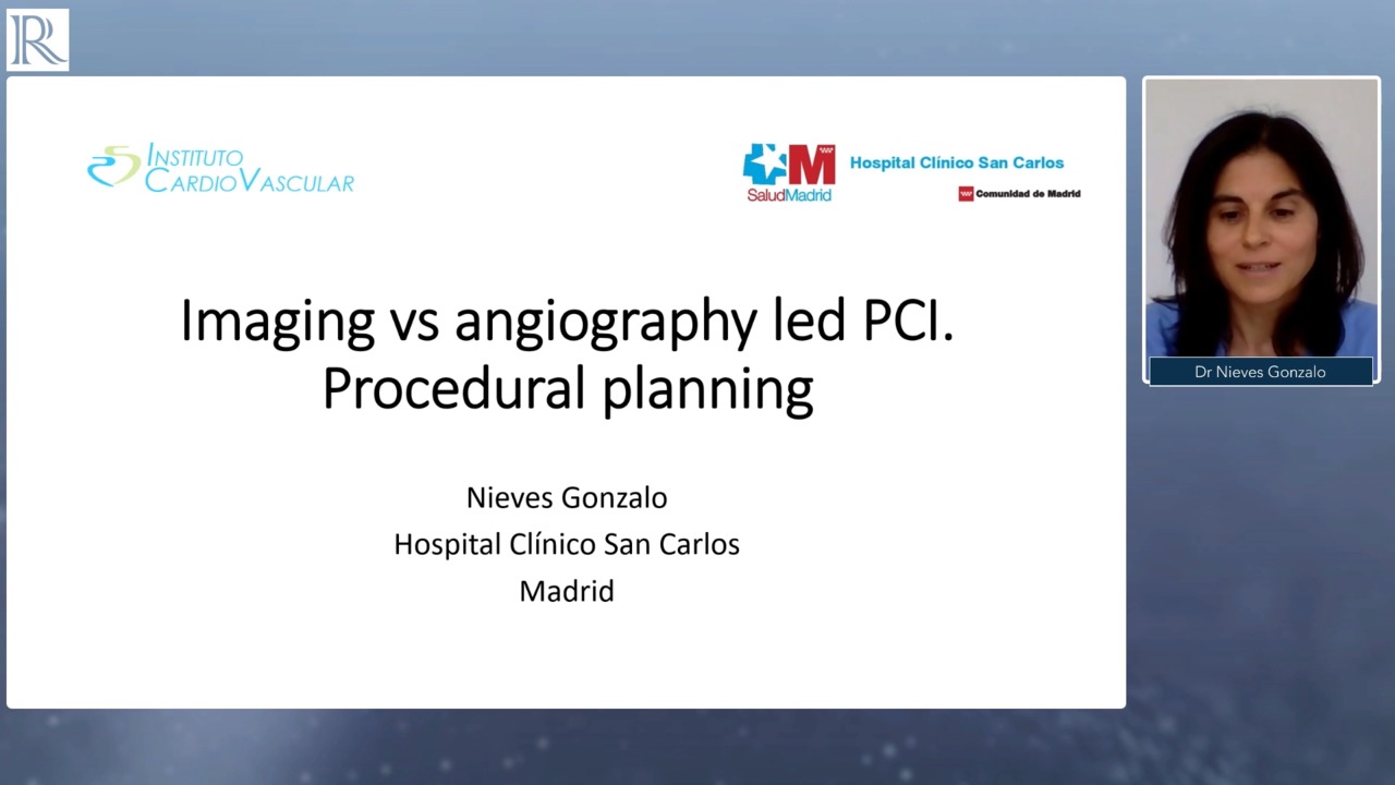 15m 56sPart 2 | Session 1 Plenary and 'Use of Imaging Pre-Stent for Procedural Planning’ Presentation Evan Shlofmitz, Nieves Gonzalo, Jonathan Hill
-
 7m 44sPart 2 | Session 2 Deep Dive into Calcification and Plaque Modification’ Presentation Jonathan Hill
-
 8m 29sPart 2 | Session 3 Proceed with ‘PCI Optimisation’ Presentation Evan Shlofmitz
-
 13m 43sPart 2 | Session 4 'Use of Imaging Pre-Stent for Procedural Planning’ - Case Study Nieves Gonzalo
-
 10m 39sPart 2 | Session 5 Proceed with ‘PCI Optimisation’ - Case Study Evan Shlofmitz
-
 3mPart 2 | Session 6 Faculty Discussion Evan Shlofmitz, Nieves Gonzalo, Jonathan Hill
-
 11m 45sPart 3 | Session 1 Introduction and 'Image Acquisition and Interpretation Essentials’ Presentation Matthias Lutz
-
 7m 34sPart 3 | Session 2 'OCI in angiographic ambiguity' Presentation Claudia Cosgrove
-
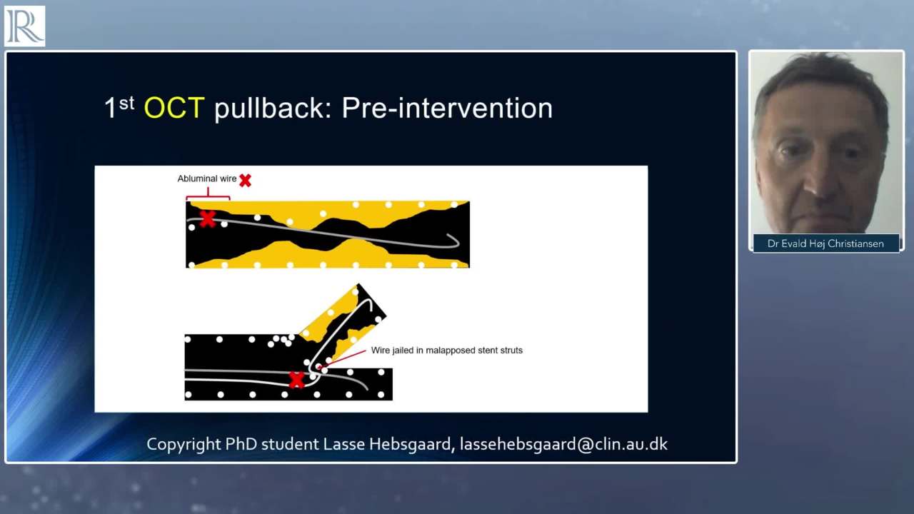 7m 34sPart 3 | Session 3 'OCI in Bifurcations and Stent Failure' Presentation Evald Høj Christiansen
-
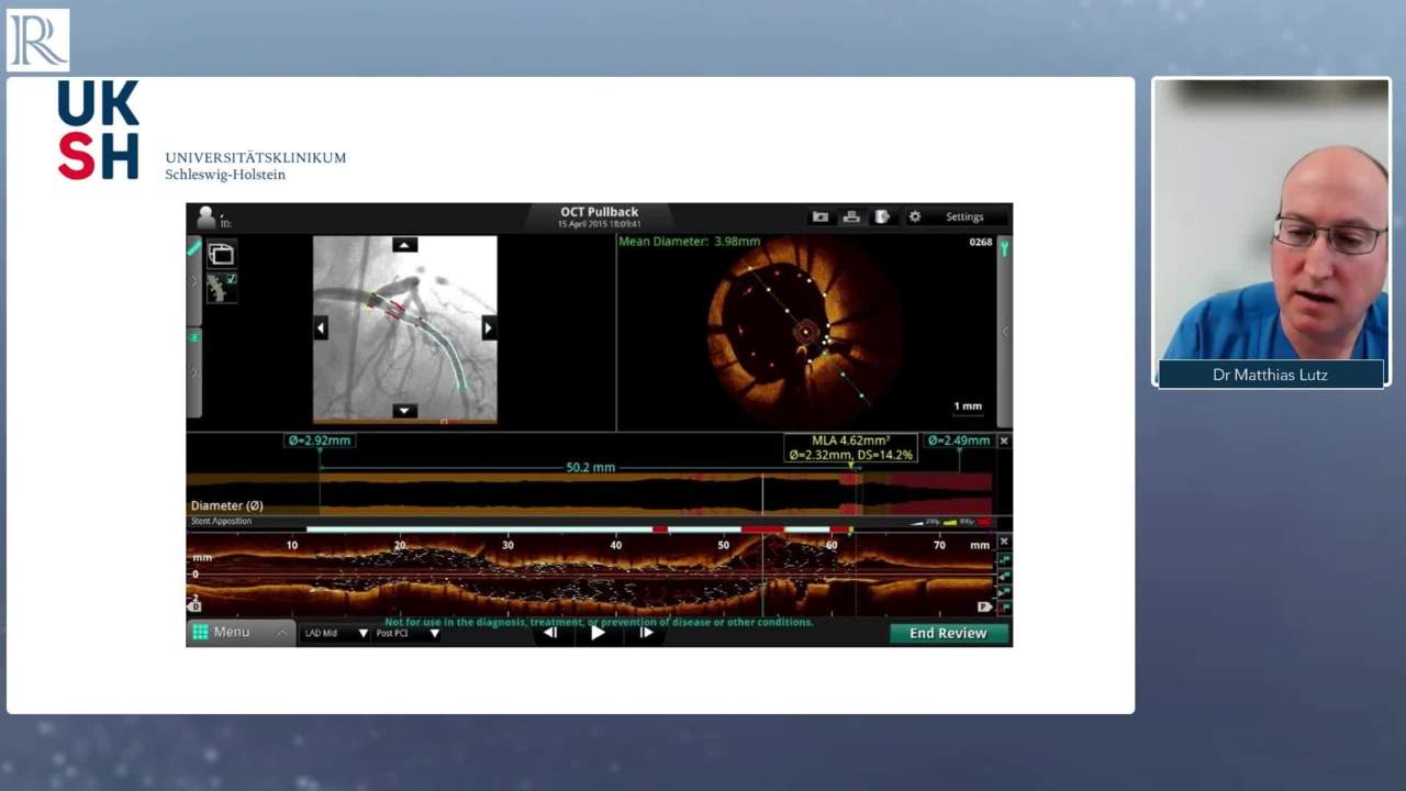
 10m 13sPart 3 | Session 4 Case study from Dr Matthias Lutz Matthias Lutz
-
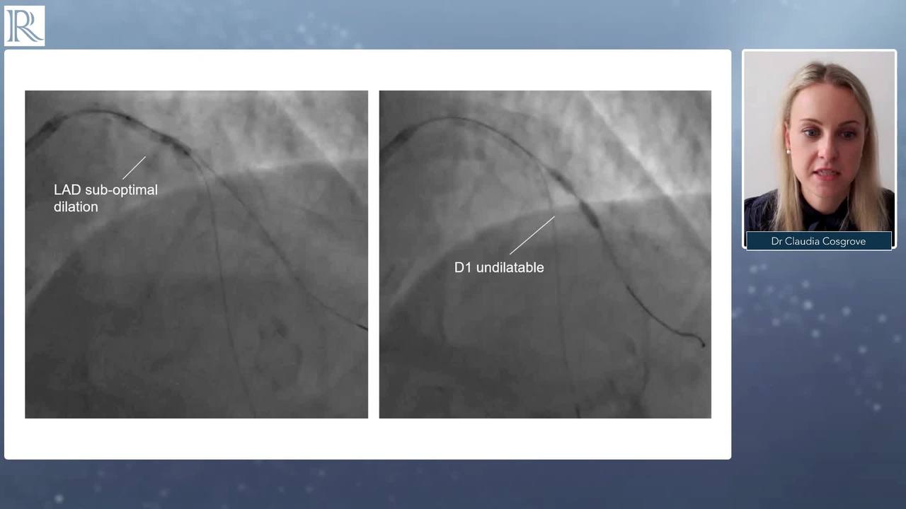
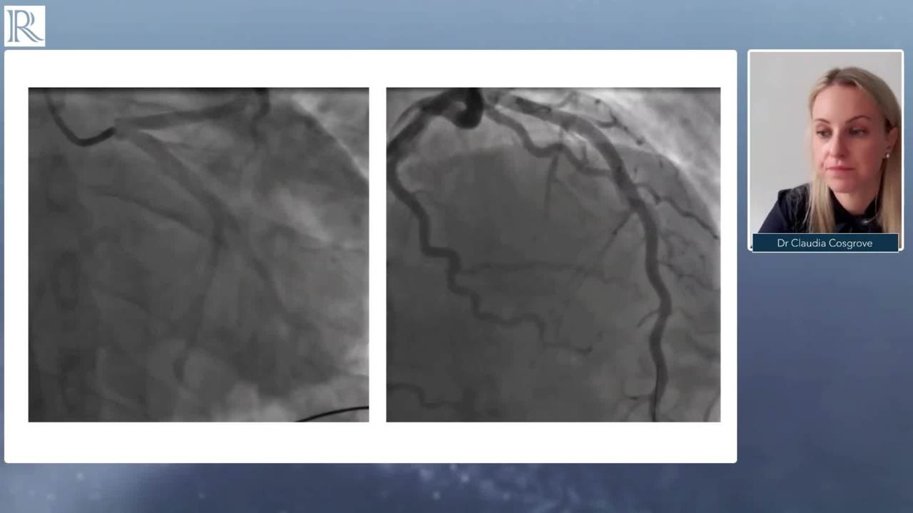 8m 48sPart 3 | Session 5 Case Study from Claudia Cosgrove Claudia Cosgrove
-
 5m 48sPart 3 | Session 6 Case Study from Evald Høj Christiansen Evald Høj Christiansen
-
 20m 7sPart 3 | Session 7 Faculty Discussion Matthias Lutz , Claudia Cosgrove, Evald Høj Christiansen
-
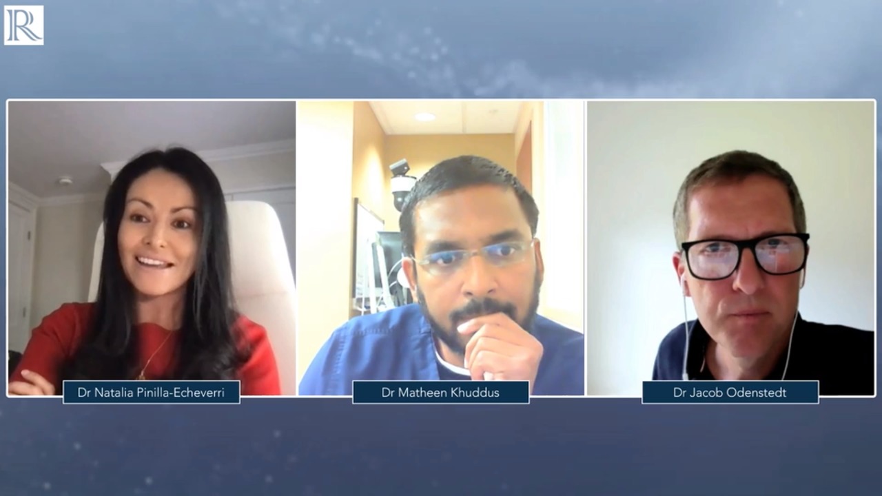
 9m 41sPart 4 | Session 1 Introduction and 'Light Lab Data' Presentation Natalia Pinilla
-
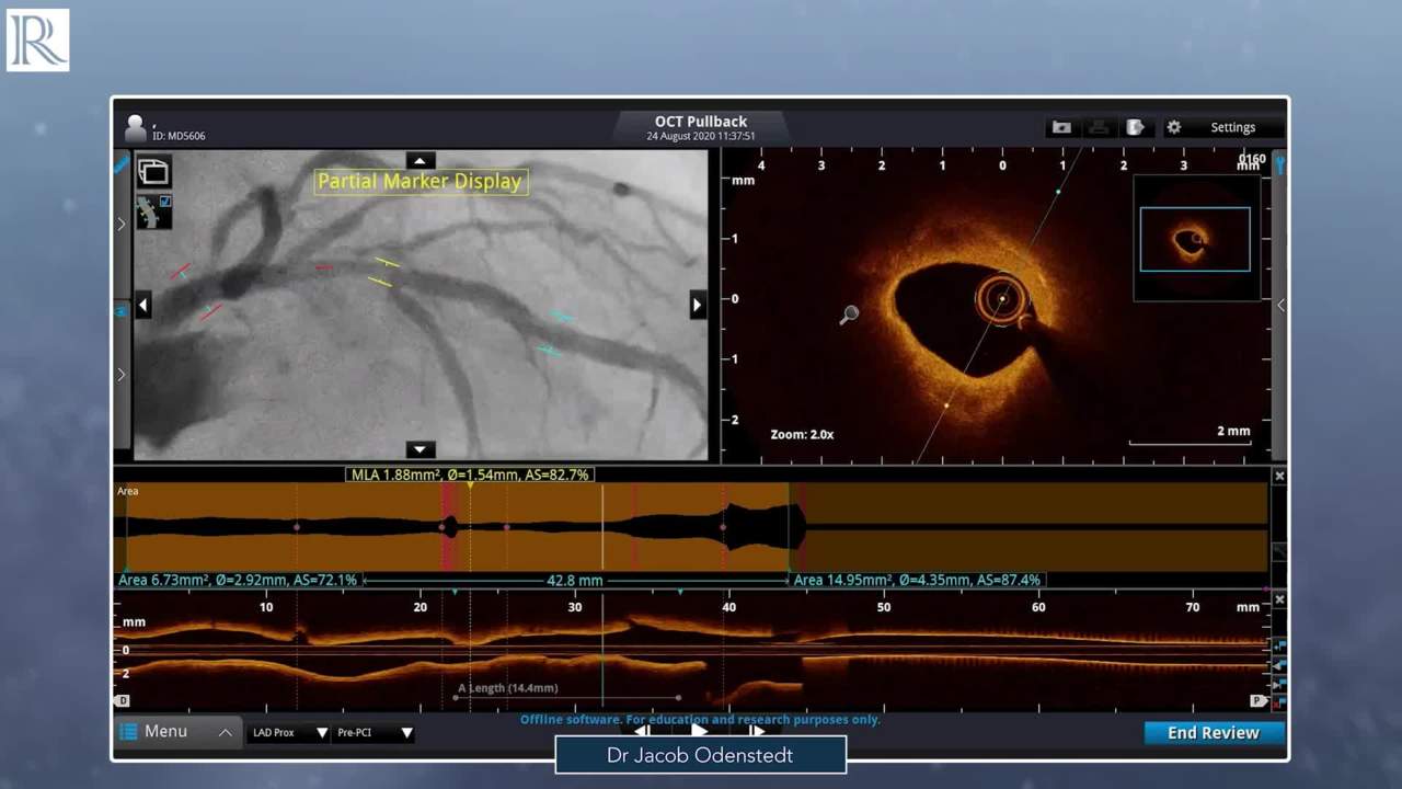
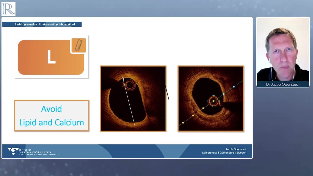 7m 56sPart 4 | Session 2 'MLD' Presentation Jacob Odenstedt
-
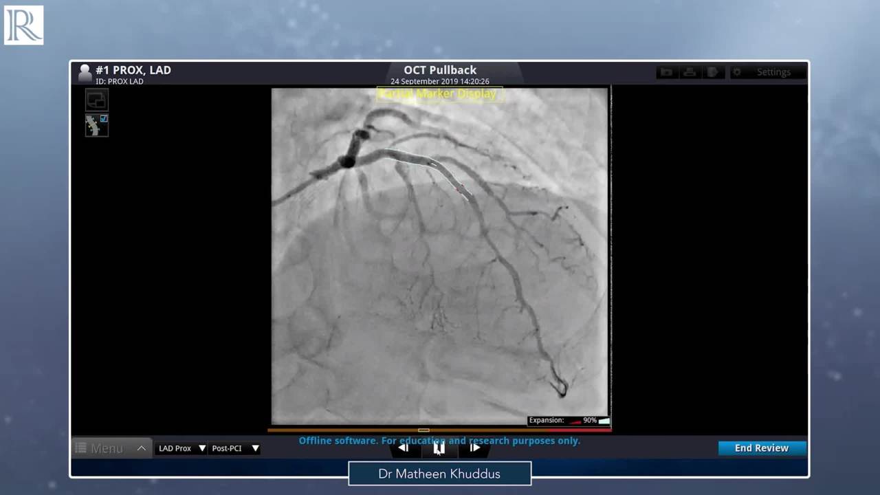
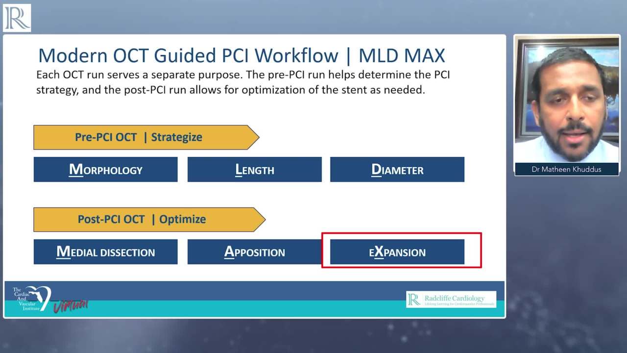 10m 2sPart 4 | Session 3 'Post PCI Optimization the MAX in MLDMAX' Presentation Matheen A Khuddus
-
 12m 7sPart 4 | Session 4 Case Study - Long Lipidic Plaque Jacob Odenstedt
-
 8m 1sPart 4 | Session 5 Case Study - Calcified Plaque Matheen A Khuddus
-
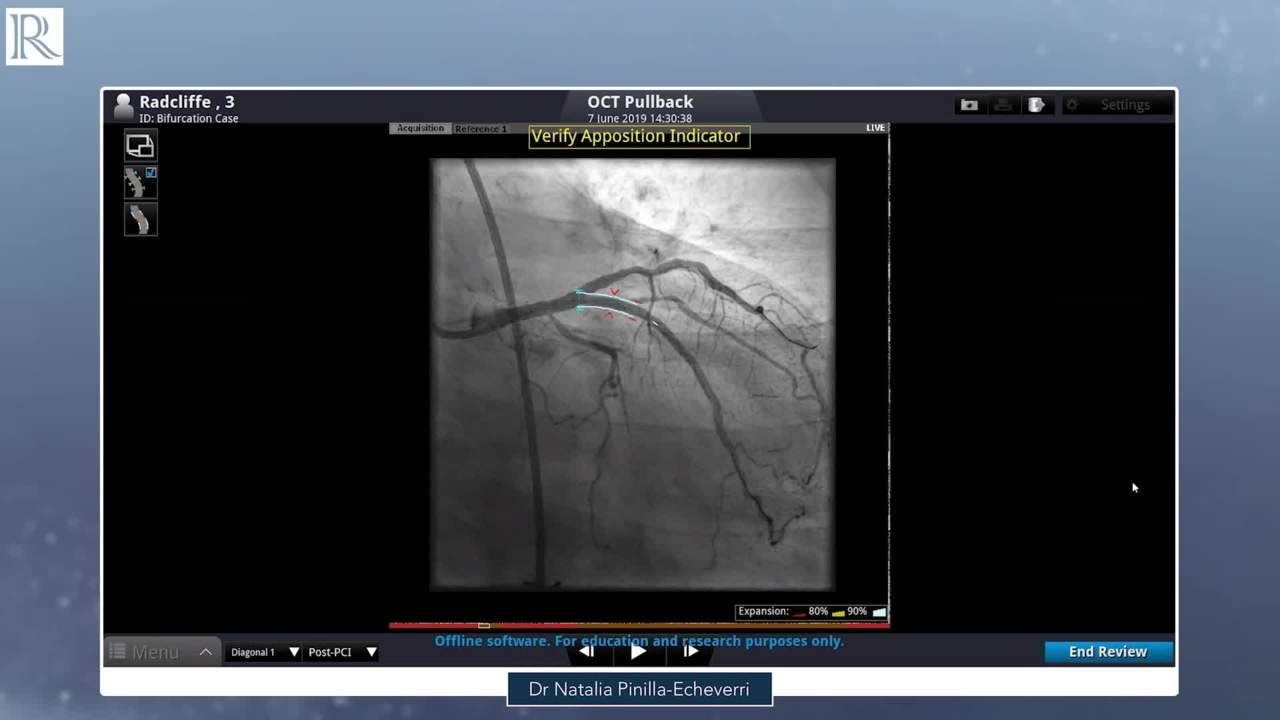 9m 44sPart 4 | Session 6 Case Study - Bifurcation Natalia Pinilla
Overview
Contemporary invasive imaging techniques improve the detection of coronary details and have great potential for improving clinical outcomes, due the lower risk of in-stent restenosis and thrombosis. This webinar series provides an overview of the current use of intravascular ultrasound (IVUS) and optical coherence tomography (OCT), the relative advantages of each technique and real-world insight from imaging experts.
This programme has been designed to offer education on proper image acquisition, interpretation, and correct decision-making to optimise the use of contemporary imaging.
Note, this on-demand version is not CME accredited.
Learning Objectives
- Incorporate contemporary imaging for PCI in appropriate patients
- Differentiate between modern imaging techniques based on clinical data
- Recall best practices for image acquisition, interpretation and decision-making
- Interpret image data and make clinical decisions generated from existing case study data
Target Audience
- Interventional cardiologists
- Physicians within the peripheral intervention space
- Imaging specialists
More from this programme
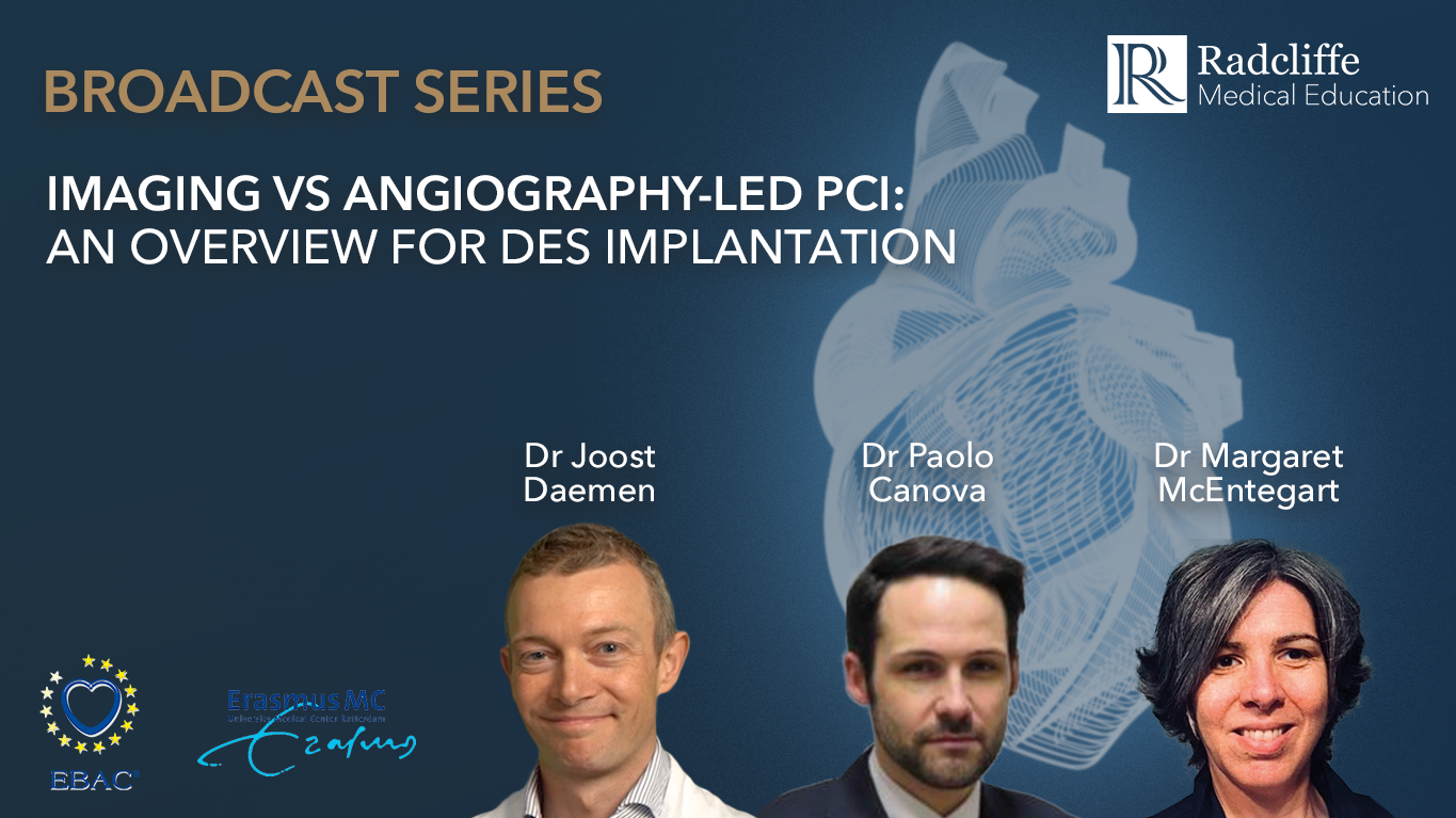
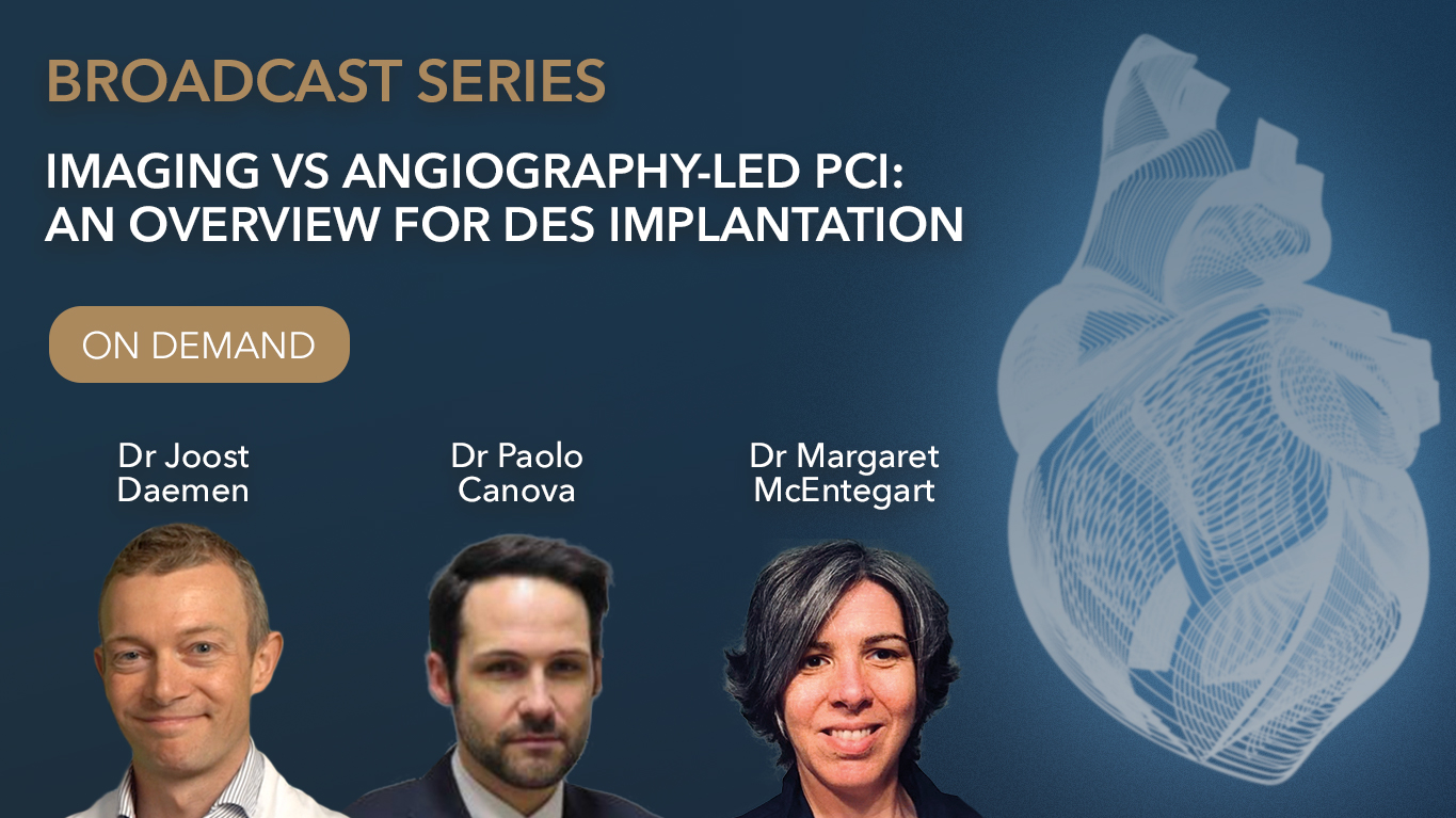
Part 1
Imaging vs angiography-led PCI: An overview for DES implantation
Part 2
Imaging vs angiography-led PCI: What does success look like?
Part 3
Intracoronary imaging for PCI: A practical approach to OCT
Part 4
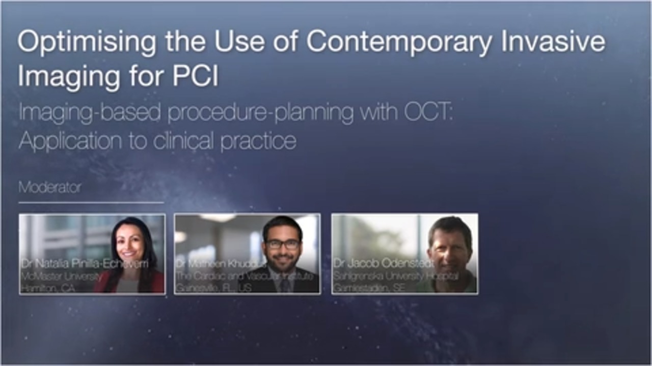
Imaging-based Procedure-planning with OCT: Application to Clinical Practice
Faculty Biographies
Natalia Pinilla-Echeverri
Dr Pinilla is originally from Colombia and received her MD from Universidad de Caldas in Manizales, Colombia. She moved to Spain to complete training in Internal Medicine and Cardiology and then moved to Canada to complete an Interventional Cardiology, non-invasive Cardiac Computed Tomography Angiography and advanced Intracoronary Imaging fellowship at McMaster University, Hamilton, Canada. She also obtained a Master’s Degree in Health Research Methodology from Universidad de Cordoba and is currently in the PhD Program in Universidad Complutense in Madrid, Spain. She is passionate about acute coronary syndrome research and leads physiology and intravascular imaging physicians and fellows training worldwide.
Jacob Odenstedt
Senior Interventional Cardiologist Sahlgrenska University Hospital Gothenburg Sweden