Article

Haemodynamics of Heart Failure With Preserved Ejection Fraction: A Clinical Perspective

Register or Login to View PDF Permissions
Permissions× For commercial reprint enquiries please contact Springer Healthcare: ReprintsWarehouse@springernature.com.

For permissions and non-commercial reprint enquiries, please visit Copyright.com to start a request.

For author reprints, please email rob.barclay@radcliffe-group.com.
Average (ratings)
No ratings
Your rating

Abstract

Despite the burden of heart failure (HF) with preserved ejection fraction (HFpEF), its pathophysiological mechanisms remain controversial and are likely to be multifactorial. Indeed, it has been suggested that HFpEF may represent “a forest of a variety of trees”, because of heterogeneity in pathophysiological mechanisms involved and phenotypic expression of the disease. A better understanding of HFpEF is crucial for the development of appropriate therapeutic targets. Recent studies on HFpEF have highlighted its particular haemodynamic features, and haemodynamic derangements are critical to both early and advanced stages of the disease. By definition, haemodynamic properties are determined by the result of the dynamic interchange between the heart, vasculature, and autonomic nervous system, which regulate the circulation of blood. Importantly, it has been shown that both intrinsic and extrinsic factors are implicated in the haemodynamic impairments typical of HFpEF patients. Thus, understanding of HFpEF haemodynamics requires consideration of the interplay between both cardiac and non-cardiac factors.

Disclosure:The authors have no conflicts of interest to declare.

Received:

Accepted:

Correspondence Details:Michele Senni, MD, Cardiology, Heart Failure and Heart Transplant Unit, Azienda Ospedaliera Papa Giovanni XXIII, Bergamo, Italy. E: msenni@hpg23.it

Copyright Statement:

The copyright in this work belongs to Radcliffe Medical Media. Only articles clearly marked with the CC BY-NC logo are published with the Creative Commons by Attribution Licence. The CC BY-NC option was not available for Radcliffe journals before 1 January 2019. Articles marked ‘Open Access’ but not marked ‘CC BY-NC’ are made freely accessible at the time of publication but are subject to standard copyright law regarding reproduction and distribution. Permission is required for reuse of this content.

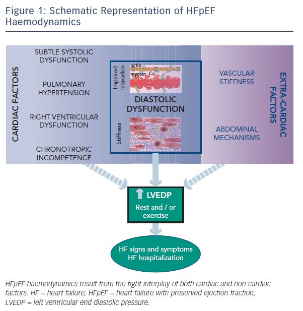
Despite the burden of heart failure (HF) with preserved ejection fraction (HFpEF),1 its pathophysiological mechanisms remain controversial and are likely to be multifactorial.2,3,4 The lack of a comprehensive paradigm applicable to all patients suggests that haemodynamic derangements responsible for this disorder may be quite heterogeneous. As recently highlighted, haemodynamic features of HFpEF involve both cardiac and extra-cardiac mechanisms. Studies on HFpEF have shown diastolic abnormalities, subtle systolic dysfunction, pulmonary hypertension, right ventricular dysfunction and chronotropic incompetence, in addition to ventricular–vascular mechanisms and abdominal factors. In this short review we highlight and discuss the different mechanisms characterizing HFpEF haemodynamics, which finally lead to elevated left ventricular end diastolic pressure (LVEDP), a common hallmark of this multifaceted syndrome (see Figure 1).

Cardiac Factors in HFpEF Haemodynamics

Patients with HFpEF are considered to be predominantly elderly women with hypertension, left atrial enlargement, obesity, and with specific pathophysiological abnormalities in cardiac structure such as myocyte hypertrophy, interstitial fibrosis, inflammation, and microvascular dysfunction. Thus, it has been postulated that the signs and symptoms of HF may be primarily a consequence of progressive abnormalities in diastolic function domains.5 The importance of diastolic left ventricular dysfunction in HFpEF has been confirmed by the majority of invasive and non-invasive haemodynamic studies, which show uniform presence at rest of slow active left ventricle (LV) relaxation and elevated passive LV stiffness.5,6,7,8 Furthermore, it has been demonstrated that elevated diastolic LV stiffness may limit cardiac performance during exercise-associated tachycardia or rapid pacing.9 In contrast, Kawaguchi et al, using the multi-beat conductance catheter technique, demonstrated in a small group of patients and controls that ventricular-arterial stiffening was the predominant mechanism underlying elevated LVEDP. However, they did not show differences in relaxation and stiffness in subjects with HFpEF compared with HF-free controls.10 Therefore, other intrinsic mechanisms have been advocated in HFpEF haemodynamics, such as subtle systolic dysfunction, pulmonary hypertension and right ventricular (RV) dysfunction.

While left ventricular ejection fraction (LVEF) is the most widely used index of systolic function, applied to distinguish HFpEF from mid-range and reduced LVEF HF patients,11 it is dependent on loading conditions and chamber size. Therefore, it is a poor measure of contractility. Importantly, it has been shown that HFpEF patients – despite the preserved LVEF – have subtle systolic dysfunction at rest, by means of reduced LV strain at echocardiographic imaging, and this dysfunction has prognostic relevance.12,13 Furthermore, it has been suggested that contractile dysfunction may contribute to inadequate myocardial response to efforts, leading to the appearance and aggravation of HF symptoms.14,15 Indeed, a recent study in HFpEF subjects examined cardiac systolic reserve during exercise and found that contractility increases were depressed.16 Therefore, the exercise test may unmask mild deficits in systolic function in HFpEF.

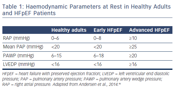
Previous studies have also reported a high prevalence of pulmonary hypertension (PH) in HFpEF.17 Importantly, PH portends worse outcome in HFpEF patients.18,19 It has been shown that the degree of PH is similar in subjects with HFpEF and HF with reduced ejection fraction (HFrEF), and is largely reversible with acute infusion of sodium nitroprusside. However, the combined potentially greater risk of hypotension or depression of stroke volume suggests that vasodilator-based approaches may not be as broadly applicable to HFpEF as they are to HFrEF.20 Another study has shown that pulmonary artery systolic pressure (PASP) rises along with pulmonary artery capillary wedge pressure (PAWP) in patients with both hypertension and HFpEF.18 However, PASP remains higher in HFpEF, even when adjusting for PAWP, suggesting a pre-capillary component to PH on top of pulmonary venous hypertension (PVH).18 Distinguishing these factors may be difficult. By definition, an elevated PAWP (i.e. >15 mmHg) characterises PVH, while pulmonary arterial hypertension (PAH) is typically associated with a normal PAWP. Of note, estimation of PAWP by non-invasive methods is suboptimal. This is because the most used echocardiographic index of elevated PAWP – E/E’ – has been shown to be only modestly correlated with supine PAWP in patients with unexplained dyspnoea and preserved LVEF and, in general, this index lacks sensitivity.21,22,23 Therefore invasive evaluation currently provides more reliable diagnostic information (see Table 1). Of note, PAWP obtained at the time of right heart catheterisation is influenced by resting conditions and the patient’s volume status at the time of the procedure. Indeed, it has been demonstrated that resting haemodynamics may be normal, while performing right heart catheterisation during supine exercise may unmask a diagnosis of HFpEF.24 Furthermore, invasive exercise testing may substantially improve prediction of long-term mortality in patients with suspected HFpEF.25 Because this technique is mainly used in research institutions, a possible alternative is to perform a fluid challenge at the time of catheterisation, to confirm HFpEF presence,26 and eventually to differentiate PAH from PVH. In fact, a recent study showed that among 207 patients with a suspected diagnosis of PAH, one fifth developed elevated PAWP after a fluid bolus, and were reclassified as having PVH.27 Nonetheless, it is important to remember that approximately 20 % of normal adults may develop PAWP >15 mmHg with acute saline infusion.28 Overall, such data imply that many patients with PH may have an under-recognised component of PVH linked to left-sided HF, which is manifested more under conditions of exertion or volume loading.29 In particular, exercise stress testing or volume challenge are indicated at the time of invasive procedure for patients presenting with PH and normal PAWP who are obese and/or who have a dilated left atrium.30 However, there is not presently enough evidence on standardisation of these procedures. There are no validated cut-off values for a pathological haemodynamic response during exercise or after acute saline infusion that would allow a clear classification of HFpEF. Generating these values by performing diagnostic trials is of major importance, but at present it is difficult to recommend routine exercise haemodynamic testing and/or a volume challenge for clinical practice.

Another invasive haemodynamic study has recently shown that RV dysfunction is common in HFpEF and is caused by both RV contractile impairment and afterload mismatch from PH.31 In this study, the factors associated with RV dysfunction were increasing pulmonary artery pressures, atrial fibrillation, male sex and LV dysfunction. It has also been demonstrated that patients with HFpEF display impaired RV reserve during exercise that is associated with high filling pressures and inadequate cardiac output responses.32 These findings highlight the importance of biventricular dysfunction in HFpEF haemodynamics.

Figure 1: Schematic Representation of HFpEF Haemodynamics

Article image

Table 1: Haemodynamic Parameters at Rest in Healthy Adults and HFpEF Patients

Article image

Chronotropic incompetence represents another important characteristic of HFpEF, which has been described in approximately 30 % of patients.33,34,35 Indeed, chronotropic reserve is depressed in HFpEF even when compared with older, age-matched controls, independently from rate-lowering medication use. Chronotropic incompetence may help to partially explain why most patients with HFpEF complain of symptoms only during physical exertion. Since the increase in plasma catecholamine with exercise is similar in HFpEF and healthy controls, it has been suggested that chronotropic incompetence may be related to deficits in beta-adrenergic stimulation.33 Additionally, autonomic dysfunction may be a contributing factor, as heart rate recovery is abnormal and baroreflex sensitivity impaired in HFpEF.34

Extrinsic Factors in HFpEF Haemodynamics

An alternative model for HF development in HFpEF patients underscores the contribution of non-myocardial factors to systolic and diastolic LV performance.

Cardiac function is affected by the net balance between afterload and preload.36 Central aortic stiffness, increasing systolic load and negatively affecting ventricular–vascular coupling, may accelerate HF development in at-risk patients. Of note, aortic stiffness increases with age, particularly in women with hypertension, and is a precursor of incident HF.37,38 To preserve adequate coupling among the heart and the arterial system, ventricular systolic stiffening also increases, and this combined ventricular–vascular stiffening is a hallmark of HFpEF.10,39 This limits LV systolic reserve, increases the cardiac energy demands required to enhance cardiac output, and plays a central role in arterial pressure liability accompanying small changes in LV preload.10

Figure 2: Differences in Left Ventricular and Left Atrial Pressure Recordings

Article image

Other extrinsic factors advocated in HFpEF haemodynamics comprise abdominal mechanisms. In many HFpEF patients fluid retention may occur in the abdominal cavity, with bowel congestion leading to endotoxin translocation and systemic inflammation. In the same way, systemic inflammation may also be induced by comorbidities such as obesity, diabetes mellitus or chronic obstructive pulmonary disease that are highly prevalent in these patients, and this has been suggested as a possible cause of myocardial structural and functional alterations.40,41 Of note, the increased neurohormonal activation typical of HF results in venoconstriction, with impaired capacitance function of the splanchnic vasculature. Increased capillary hydrostatic pressure leads to a rise in intra-abdominal pressure and eventually also to organ dysfunction. Indeed, HFpEF is frequently associated with renal impairment, as chronic kidney disease (CKD) occurs in up to two thirds of HFpEF patients and is associated with poor prognosis.42,43,44 There is a bidirectional link between HFpEF and CKD, as it has been shown that venous congestion may lead to CKD and, vice versa, renal impairment begets congestion and HF. Renal impairment causes metabolic derangements and affects circulating factors causing an activated systemic inflammatory state and endothelial dysfunction. This may lead to hypertrophy, myocardial stiffening, and interstitial fibrosis via cross-talk between the endothelium and cardiomyocyte, finally causing haemodynamic impairments.45

HFpEF Haemodynamics: Critical Appraisal

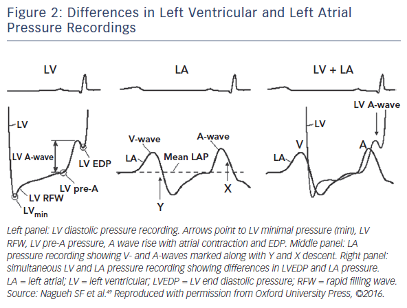
While diastolic dysfunction was originally considered pathognomonic in HFpEF haemodynamics,5,6,7,8,9 abnormalities of active LV relaxation and/or passive stiffness have been described by most but not all invasive haemodynamic studies.10 There are different reasons for these discrepancies, such as heterogeneity in study populations, differences in control groups, the use of invasive or non-invasive studies, or examinations being performed only at rest or also during effort. Additionally, a major challenge to the field is that truly representative experimental models of HFpEF do not exist, and human data – particularly direct myocardial analysis – remain limited, with very small populations having been studied. There are no data from beating muscle or cells from human hearts, and existing animal models fail to capture the complexity of the human disease. Furthermore, it should be emphasised that only left heart catheterisation allows for direct measurement of LVEDP, as well as the kinetics of relaxation and passive chamber stiffness through pressure-volume recordings (PV loops). However, these assessments require highfidelity micromanometer and conductance catheter systems, which are demanding techniques that are not easily reproducible and not widely available in clinical settings. Of note, even though it has been suggested that a high-quality PAWP tracing is just as robust as directly measured LVEDP,46 this assumption is incorrect in the case of LV disease. This is because a strong atrial contribution to LV filling can occur in this condition, which can translate in a LVEDP considerably higher than the mean left atrial pressure and PAWP (see Figure 2).47 Overall, these considerations seem to suggest that diastolic dysfunction may still have a central role in HFpEF haemodynamics, although it may be demanding to prove it in clinical and experimental studies (see Figure 1).

Finally, the recognition of diastolic LV dysfunction as the predominant mechanism underlying HFpEF haemodynamics does not necessarily imply that the latter represents the sole contributor to haemodynamic derangements. Indeed, numerous other mechanisms have been recently identified and may play important roles. Among these, both cardiac and extra-cardiac factors should be assessed to adequately interpret HFpEF haemodynamics. However, it should be emphasised that ultimately most of these mechanisms negatively affect LVEDP, which represents the hallmark of HFpEF, together with normal LV dimensions and preserved LVEF. Raised LVEDP justifies HF signs and symptoms, may lead to myocardial ischemia, fibrosis, and structural impairment, and portends adverse prognosis in HFpEF patients. An approach centred on the specific pathophysiological abnormalities in cardiac structure and function, in particular diastolic dysfunction, may be more effective for therapeutic discovery.48

References

  1. Owan TE, Hodge DO, Herges RM, et al. Trends in prevalence and outcome of heart failure with preserved ejection fraction. N Engl J Med 2006;355:251–9.
    Crossref | PubMed
  2. Senni M, Paulus WJ, Gavazzi A, et al. New strategies for heart failure with preserved ejection fraction: the importance of targeted therapies for heart failure phenotypes. Eur Heart J 2014;35:2797–815.
    Crossref | PubMed
  3. D’Elia E, Vaduganathan M, Gori M, et al. Role of biomarkers in cardiac structure phenotyping in heart failure with preserved ejection fraction: critical appraisal and practical use. Eur J Heart Fail 2015;17:1231–9.
    Crossref | PubMed
  4. Shah AM, Pfeffer MA. The many faces of heart failure with preserved ejection fraction. Nat Rev Cardiol 2012;9:555–6.
    Crossref | PubMed
  5. Zile MR, Baicu CF, Gaasch WH. Diastolic heart failure— abnormalities in active relaxation and passive stiffness of the left ventricle. N Engl J Med 2004;350:1953–9.
    Crossref | PubMed
  6. Phan TT, Abozguia K, Nallur Shivu G, et al. Heart failure with preserved ejection fraction is characterized by dynamic impairment of active relaxation and contraction of the left ventricle on exercise and associated with myocardial energy deficiency. J Am Coll Cardiol 2009;54:402–9.
    Crossref | PubMed
  7. Penicka M, Bartunek J, Trakalova H, et al. Heart failure with preserved ejection fraction in outpatients with unexplained dyspnea: a pressure-volume loop analysis. J Am Coll Cardiol 2010;55:1701–10.
    Crossref | PubMed
  8. Prasad A, Hastings JL, Shibata S, et al. Characterization of static and dynamic left ventricular diastolic function in patients with heart failure with a preserved ejection fraction. Circ Heart Fail 2010;3:617–26.
    Crossref | PubMed
  9. Westermann D, Kasner M, Steendijk P, et al. Role of left ventricular stiffness in heart failure with normal ejection fraction. Circulation 2008;117:2051–60.
    Crossref | PubMed
  10. Kawaguchi M, Hay I, Fetics B, et al. Combined ventricular systolic and arterial stiffening in patients with heart failure and preserved ejection fraction: implications for systolic and diastolic reserve limitations. Circulation 2003;107:714–20.
    Crossref | PubMed
  11. Ponikowski P, Voors AA, Anker SD, et al. 2016 ESC Guidelines for the diagnosis and treatment of acute and chronic heart failure: The Task Force for the diagnosis and treatment of acute and chronic heart failure of the European Society of Cardiology (ESC) Developed with the special contribution of the Heart Failure Association (HFA) of the ESC. Eur Heart J 2016;37:2129–200.
    Crossref | PubMed
  12. Kraigher-Krainer E, Shah AM, Gupta DK, et al. Impaired systolic function by strain imaging in heart failure with preserved ejection fraction. J Am Coll Cardiol 2014; 63:447–56.
    Crossref | PubMed
  13. Shah AM, Claggett B, Sweitzer NK, et al. Prognostic importance of impaired systolic function in heart failure with preserved ejection fraction and the impact of spironolactone. Circulation 2015;132:402–14.
    Crossref | PubMed
  14. Borlaug BA, Olson TP, Lam CS, et al. Global cardiovascular reserve dysfunction in heart failure with preserved ejection fraction. J Am Coll Cardiol 2010;56:845–54.
    Crossref | PubMed
  15. Maeder MT, Thompson BR, Brunner-La Rocca HP, et al. Hemodynamic basis of exercise limitation in patients with heart failure and normal ejection fraction. J Am Coll Cardiol 2010;56:855–63.
    Crossref | PubMed
  16. Abudiab MM, Redfield MM, Melenovsky V, et al. Cardiac output response to exercise in relation to metabolic demand in heart failure with preserved ejection fraction. Eur J Heart Fail 2013;15:776–85.
    Crossref | PubMed
  17. Burke MA, Katz DH, Beussink L, et al. Prognostic importance of pathophysiologic markers in patients with heart failure and preserved ejection fraction. Circ Heart Fail 2014;7:288–99.
    Crossref | PubMed
  18. Lam CS, Roger VL, Rodeheffer RJ, et al. Pulmonary hypertension in heart failure with preserved ejection fraction: a community-based study. J Am Coll Cardiol 2009;53:1119–26.
    Crossref | PubMed
  19. Shah AM, Shah SJ, Anand IS, et al.; TOPCAT Investigators. Cardiac structure and function in heart failure with preserved ejection fraction: baseline findings from the echocardiographic study of the treatment of preserved cardiac function heart failure with an aldosterone antagonist trial. Circ Heart Fail. 2014;7:104–15.
    Crossref | PubMed
  20. Schwartzenberg S, Redfield MM, From AM, et al. Effects of vasodilation in heart failure with preserved or reduced ejection fraction implications of distinct pathophysiologies on response to therapy. J Am Coll Cardiol 2012;59:442–51.
    Crossref | PubMed
  21. Santos M, Rivero J, McCullough SD, et al. E/e’ Ratio in Patients With Unexplained Dyspnea: Lack of Accuracy in Estimating Left Ventricular Filling Pressure. Circ Heart Fail. 2015;8:749–56.
    Crossref | PubMed
  22. Ommen SR, Nishimura RA, Appleton CP, et al. Clinical utility of Doppler echocardiography and tissue Doppler imaging in the estimation of left ventricular filling pressures: a comparative simultaneous Doppler-catheterization study. Circulation 2000;102:1788–94.
    Crossref | PubMed
  23. From AM, Lam CS, Pitta SR, et al. Bedside assessment of cardiac hemodynamics: the impact of noninvasive testing and examiner experience. Am J Med 2011;124:1051–7.
    Crossref | PubMed
  24. Borlaug BA, Nishimura RA, Sorajja P, et al. Exercise hemodynamics enhance diagnosis of early heart failure with preserved ejection fraction. Circ Heart Fail 2010;3:588–95.
    Crossref | PubMed
  25. Dorfs S, Zeh W, Hochholzer W, et al. Pulmonary capillary wedge pressure during exercise and long-term mortality in patients with suspected heart failure with preserved ejection fraction. Eur Heart J 2014;35:3103–12.
    Crossref | PubMed
  26. Fujimoto N, Borlaug BA, Lewis GD, et al. Hemodynamic responses to rapid saline loading: the impact of age, sex, and heart failure. Circulation 2013;127:55–62.
    Crossref | PubMed
  27. Robbins IM, Hemnes AR, Pugh ME, et al. High prevalence of occult pulmonary venous hypertension revealed by fluid challenge in pulmonary hypertension. Circ Heart Fail 2014;7:116–22.
    Crossref | PubMed
  28. Andersen MJ, Olson TP, Melenovsky V, et al. Differential hemodynamic effects of exercise and volume expansion in people with and without heart failure. Circ Heart Fail 2015;8:41–8.
    Crossref | PubMed
  29. Borlaug BA. Invasive assessment of pulmonary hypertension: time for a more fluid approach? Circ Heart Fail 2014;7:2–4.
    Crossref | PubMed
  30. Maor E, Grossman E, Gingy Balmor R, et al. Exercise haemodynamics may unmask the diagnosis of diastolic dysfunction among patients with pulmonary hypertension. Eur J Heart Fail 2015;17:151–58.
    Crossref | PubMed
  31. Melenovsky V, Hwang SJ, Lin G, et al. Right heart dysfunction in heart failure with preserved ejection fraction. Eur Heart J 2014;35:3452–62.
    Crossref | PubMed
  32. Borlaug BA, Kane GC, Melenovsky V, et al. Abnormal right ventricular-pulmonary artery coupling with exercise in heart failure with preserved ejection fraction. Eur Heart J 2016; epub ahead of press.
    Crossref | PubMed
  33. Borlaug BA, Melenovsky V, Russell SD, et al. Impaired chronotropic and vasodilator reserves limit exercise capacity in patients with heart failure and a preserved ejection fraction. Circulation 2006;114:2138–47.
    Crossref | PubMed
  34. Phan TT, Nallur Shivu G, Abozguia K, et al. Impaired heart rate recovery and chronotropic incompetence in patients with heart failure with preserved ejection fraction. Circ Heart Fail 2010;3:29–34.
    Crossref | PubMed
  35. Brubaker PH, Joo KC, Stewart KP, et al. Chronotropic incompetence and its contribution to exercise intolerance in older heart failure patients. J Cardiopulm Rehabil 2006;26:86–9.
    Crossref | PubMed
  36. Borlaug BA, Kass DA. Ventricular-vascular interaction in heart failure. Heart Fail Clin 2008;4:23–36.
    Crossref | PubMed
  37. Desai AS, Mitchell GF, Fang JC, et al. Central aortic stiffness is increased in patients with heart failure and preserved ejection fraction. J Card Fail 2009;15: 658–64.
    Crossref | PubMed
  38. Chantler PD, Lakatta EG, Najjar SS. Arterial-ventricular coupling: mechanistic insights into cardiovascular performance at rest and during exercise. J Appl Physiol 2008;105:42–1351.
    Crossref | PubMed
  39. Lam CS, Roger VL, Rodeheffer RJ, et al. Cardiac structure and ventricular-vascular function in persons with heart failure and preserved ejection fraction from Olmsted County, Minnesota. Circulation 2007;115:1982–90.
    Crossref | PubMed
  40. Verbrugge FH, Dupont M, Steels P, et al. Abdominal contributions to cardiorenal dysfunction in congestive heart failure. J Am Coll Cardiol 2013;62:485–95.
    Crossref | PubMed
  41. Paulus WJ, Tschöpe C. A Novel Paradigm for Heart Failure with Preserved Ejection Fraction: Comorbidities Drive Myocardial Dysfunction and Remodeling Through Coronary Microvascular Endothelial Inflammation. J Am Coll Cardiol 2013;62:263–71.
    Crossref | PubMed
  42. Gori M, Senni M, Gupta DK, et al; PARAMOUNT Investigators. Association between renal function and cardiovascular structure and function in heart failure with preserved ejection fraction. Eur Heart J 2014;35:3442–51.
    Crossref | PubMed
  43. Rusinaru D, Buiciuc O, Houpe D, et al. Renal function and long-term survival after hospital discharge in heart failure with preserved ejection fraction. Int J Cardiol 2011;147:278–82.
    Crossref | PubMed
  44. Brouwers FP, de Boer RA, van der Harst P, et al. Incidence and epidemiology of new onset heart failure with preserved vs. reduced ejection fraction in a community based cohort: 11-year follow-up of PREVEND. Eur Heart J 2013;34:1424–31.
    Crossref | PubMed
  45. Ter Maaten JM, Damman K, Verhaar MC, et al. Connecting heart failure with preserved ejection fraction and renal dysfunction: the role of endothelial dysfunction and inflammation. Eur J Heart Fail 2016;18:588–98.
    Crossref | PubMed
  46. Andersen MJ, Borlaug BA. Invasive hemodynamic characterization of heart failure with preserved ejection fraction. Heart Fail Clin 2014;10:435–44.
    Crossref | PubMed
  47. Peverill RE. “Left ventricular filling pressure(s)” - Ambiguous and misleading terminology, best abandoned. Int J Cardiol 2015;191:110–3.
    Crossref | PubMed
  48. Senni M, Gavazzi A, Gheorghiade M, et al. Heart failure at the crossroads: moving beyond blaming stakeholders to targeting the heart. Eur J Heart Fail 2015;17:760–3.
    Crossref | PubMed
  49. Nagueh SF, Smiseth OA, Appleton CP, et al; Houston, Texas; Oslo, Norway; Phoenix, Arizona; Nashville, Tennessee; Hamilton, Ontario, Canada; Uppsala, Sweden; Ghent and Liège, Belgium; Cleveland, Ohio; Novara, Italy; Rochester, Minnesota; Bucharest, Romania; and St. Louis, Missouri. Recommendations for the Evaluation of Left Ventricular Diastolic Function by Echocardiography: An Update from the American Society of Echocardiography and the European Association of Cardiovascular Imaging. Eur Heart J Cardiovasc Imaging 2016; epub ahead of press.
    Crossref | PubMed