Article

Fractional Flow Reserve –­ Can it Improve Physician Decision Making in Coronary Artery Bypass Grafting?

Register or Login to View PDF Permissions
Permissions× For commercial reprint enquiries please contact Springer Healthcare: ReprintsWarehouse@springernature.com.

For permissions and non-commercial reprint enquiries, please visit Copyright.com to start a request.

For author reprints, please email rob.barclay@radcliffe-group.com.
Average (ratings)
No ratings
Your rating

Received:

Accepted:

Citation: RadcliffeCardiology.com, July 2014

Support:The publication of this information was supported by St. Jude Medical.

Copyright Statement:

The copyright in this work belongs to Radcliffe Medical Media. Only articles clearly marked with the CC BY-NC logo are published with the Creative Commons by Attribution Licence. The CC BY-NC option was not available for Radcliffe journals before 1 January 2019. Articles marked ‘Open Access’ but not marked ‘CC BY-NC’ are made freely accessible at the time of publication but are subject to standard copyright law regarding reproduction and distribution. Permission is required for reuse of this content.

The diagnosis and treatment strategies for coronary artery disease are traditionally based on the percentage of coronary angiographic vessel stenosis. We are witnessing a gradual transition from angiographic evaluation of individual coronary artery lesions towards the combination of anatomy and physiology to determining its physiological consequences. With the introduction and rapid evolution of fractional flow reserve (FFR) technology, a new gold standard has been developed to invasively assess the physiological severity of a coronary artery stenosis.1–3 Fractional flow reserve provides a real-time measurement of the extent to which a given epicardial stenosis limits maximal myocardial flow and identifies lesions that should be corrected by revascularisation.

The Impact of FFR Technology on Revascularisation Strategies

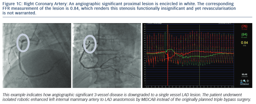
Various trials helped to support the premise that percutaneous coronary interventions (PCI) should be guided more by physiological considerations and not solely by anatomic factors.4,5,7

A number of studies designed to determine the role of FFR on coronary artery bypass grafting (CABG) have been done with promising results; however, larger prospective randomised trials are needed.10 Additionally, we still do not know what the long-term effects of not grafting angiographic stenotic lesions will be on the distal myocardium.11–13

The impact of FFR technology on revascularisation strategies

Article image

Clinical Examples that Illustrate the Disparity Between Angiographic and FFR Lesion Evaluation

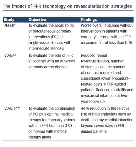
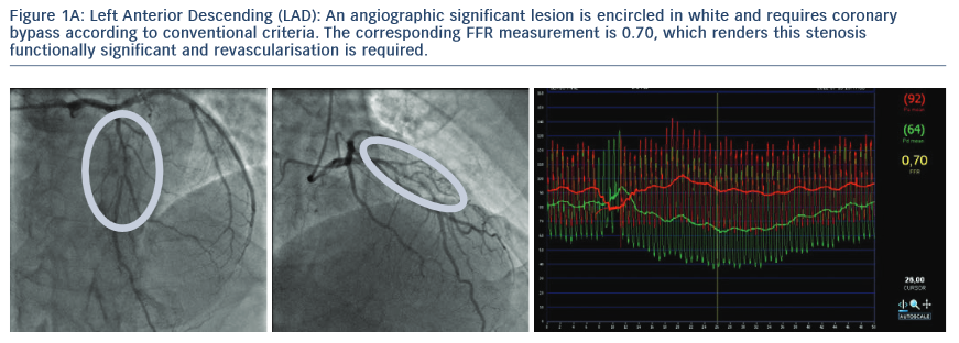
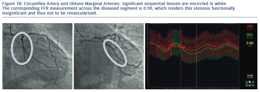
1.The Following Images Describe Significant Angiographic 3-Vessel Disease in a Patient with Stable Angina

Left Anterior Descending (LAD)

Article image

Circumflex Artery and Obtuse Marginal Arteries

Article image

Right Coronary Artery

Article image

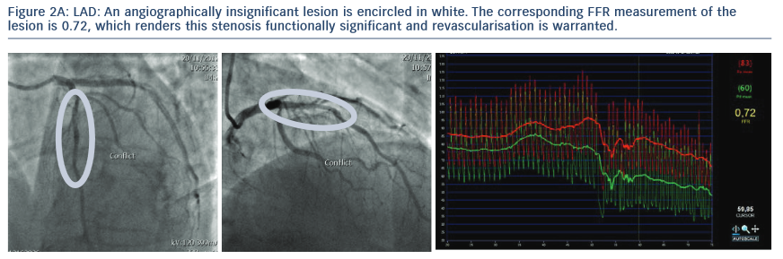
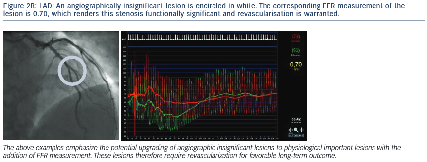
2. The Following Images Describe the Angiograms of Lesions Judged to be Potentially Insignificant in Two Separate Patients with Stable Angina

The corresponding FFR measurement of the lesion is 0.72, which renders this stenosis functionally significant and revascularisation iswarranted.

Article image

The corresponding FFR measurement of the lesion is 0.70, which renders this stenosis functionally significant and revascularisation is warranted.

Article image

Conclusion

FFR technology has proven its value in decision making about percutaneous coronary interventions but before changes are made in determining what vessels should and should not be grafted during CABG, larger prospective randomised trials with longer follow-up are needed to better understand the role of this technology in CABG. FFR-guided CABG is now under intense investigation and may have an important role in determining whether angiographic lesions should be bypassed.

Rx Only

Brief Summary: Prior to using these devices, please review the Instructions for Use for a complete listing of indications, contraindications, warnings, precautions, potential adverse events and directions for use.

Unless otherwise noted, TM indicates that the name is a trademark of, or licensed to, St. Jude Medical or one of its subsidiaries. ST. JUDE MEDICAL and the nine-squares symbol are trademarks and service marks of St. Jude Medical, Inc. and its related companies. Article courtesy of Filip Casselman MD, PhD, FETCS, Johan Van der Merwe MD, MMED (Thorax).

References

  1. Pijls N, van Son J, Kirkeeide R, De Bruyne B, Gould K. Experimental basis of determining maximum coronary, myocardial, and collateral blood flow by pressure measurements for assessing functional stenosis severity before and after percutaneous transluminal coronary angioplasty. Circulation. 1993;87,1354–67.
  2. De Bruyne B, Baudhuin T, Melin J, et al. Coronary flow reserve calculated from pressure measurements in humans. Validation with positron emission tomography. Circulation. 1994;89,1013–22.
  3. Pijls N, De Bruyne B, Peels K, et al. Measurement of fractional flow reserve to assess the functional severity of coronary-artery stenoses. The New England Journal of Medicine. 1996;334,1703–08.
  4. Pijls N, van Schaardenburgh P, Manoharan G, et al. Percutaneous coronary intervention of functionally non- significant stenosis: five-year follow-up of the DEFER Study. Journal of the American College of Cardiology. 2007;49,2105–11.
  5. Tonino P, De Bruyne B, Pijls N, et al. FAME Study Investigators. Fractional flow reserve versus angiography for guiding percutaneous coronary intervention. The New England Journal of Medicine. 2009;360,213–24.
  6. Pijls N, Fearon W, Tonino P, et al. Fractional flow reserve vs. angiography for guiding percutaneous coronary intervention in patients with multivessel coronary artery disease. Journal of the American College of Cardiology. 2010;56,177–84.
  7. De Bruyne B, Pijls N, Kalesan B, et al, FAME 2 Trial Investigators. Fractional flow reserve-guided percutaneous coronary intervention versus medical treatment in stable coronary disease. The New England Journal of Medicine. 2012;367,991–1001.
  8. De Bruyne B, Fearon W, Pijls N, et al. FAME 2 Trial Investigators. Fractional flow reserve-guided PCI for stable coronary artery disease. The New England Journal of Medicine. 2014;371,1208–1217.
  9. Hamilos M, Muller O, Cuisset T, et al. Long-term clinical outcome after fractional flow reserve-guided treatment in patients with angiographically equivocal left main coronary artery stenosis. Circulation. 2009;120,1505–12.
  10. Toth G, De Bruyne B, Casselman F, et al. (2013). Fractional flow reserve-guided versus angiography-guided coronary artery bypass graft surgery. Circulation. 2013;128,1405–11.
  11. Kubo T, Akasaka T, Shite J, et al. OCT compared with IVUS in a coronary lesion assessment: the OPUS-CLASS study. Journal of the American College of Cardiology: Cardiovascular Imaging. 2013;6,1095–1104.
  12. Bezerra H, Attizzani G, Sirbu V, et al. Optical coherence tomography versus intravascular ultrasound to evaluate coronary artery disease and percutaneous coronary intervention. Journal of the American College of Cardiology: Cardiovascular Interventions. 2013;6,228–36.
  13. Maehara A, Mintz G, Stone G. (2013). OCT versus IVUS: accuracy versus clinical utility. Journal of the American College of Cardiology: Cardiovascular Imaging. 2013;6,1105–7.