Article

Ablation of Atrial Fibrillation Drivers

Register or Login to View PDF Permissions
Permissions× For commercial reprint enquiries please contact Springer Healthcare: ReprintsWarehouse@springernature.com.

For permissions and non-commercial reprint enquiries, please visit Copyright.com to start a request.

For author reprints, please email rob.barclay@radcliffe-group.com.
Average (ratings)
No ratings
Your rating

Abstract

Pulmonary vein isolation (PVI) is central to ablation approaches for atrial fibrillation (AF), yet many patients still have arrhythmia recurrence after one or more procedures, despite evolving technologies for PVI. Ablation of localised AF drivers, which lie outside the pulmonary veins in many patients, is a practical approach that has been shown to improve success by many groups. Such localised drivers lie in atrial regions shown mechanistically to sustain AF in optical mapping and clinical studies of human AF, as well as computational and animal studies. Clinical studies now verify rotational activation by multiple mapping approaches in the same patients, at sites where ablation terminates persistent AF. This review article provides a mechanistic and clinical rationale to ablate localised drivers, and describes successful techniques for their ablation as well as pitfalls to avoid, which may explain discrepancies between results from some centres. We hope that this review will serve as a platform for future improvements in the patient-tailored ablation for complex arrhythmias.

Disclosure:This work was supported by grants from the National Institutes of Health to Sanjiv M Narayan (HL83359, HL103800). Sanjiv M Narayan is co-author of intellectual property owned by the University of California Regents and licensed to Topera Inc., and has held equity in Topera. Sanjiv M Narayan reports consulting fees and honoraria from the American College of Cardiology, Medtronic, St. Jude, Abbott and UpToDate. Paul J. Wang reports Honoraria/Consultant from Janssen, St. Jude Medical, Amgen, Medtronic; Fellowship support from Biosense Webster, Boston Scientific, Medtronic, St. Jude Medical; Clinical studies from Medtronic, Siemens, Cardiofocus, ARCA; and Stock options from Vytronus.

Received:

Accepted:

Correspondence Details:Tina Baykaner, Stanford University, 780 Welch Road MC 5773, Stanford, CA 94305, USA; E: tina4@stanford.edu

Copyright Statement:

The copyright in this work belongs to Radcliffe Medical Media. Only articles clearly marked with the CC BY-NC logo are published with the Creative Commons by Attribution Licence. The CC BY-NC option was not available for Radcliffe journals before 1 January 2019. Articles marked ‘Open Access’ but not marked ‘CC BY-NC’ are made freely accessible at the time of publication but are subject to standard copyright law regarding reproduction and distribution. Permission is required for reuse of this content.

Treatment of atrial fibrillation (AF) classically focuses on eliminating triggers near and from the pulmonary veins, which may initiate AF. However, the 1–2 year success rate of pulmonary vein isolation (PVI) remains 40–50 % for persistent AF1,2 and 50–65 % for paroxysmal AF,3–5 while supplementary linear lesions or extensive ablation at electrogram-targets have had disappointing results and may not improve the success of pulmonary vein isolation.1,2,6 In recent years, focal or rotational drivers for AF have gained increasing attention as ablation targets. This approach is now supported by wide evidence ranging from optical mapping of animal and human AF7,8 (Figure 1) to several multicentre non-randomised clinical trials of AF driver ablation, yet the paucity of randomised trials in this area still remains a shortcoming.9–14

In this review, we hope to provide an overview of the mechanistic and clinical foundation of localised drivers of human AF, technical factors explaining why they may be revealed by some but not all mapping approaches, and potential explanations for why some clinical ablation studies have been disappointing despite promising results at many independent centres.

Mechanisms of Initiation of Human AF

Triggers such as ectopic beats,15 bursts of atrial tachycardia16 or varying autonomic balance17,18 may dynamically initiate AF, despite a static atrial architecture, including fibrosis.19 Dynamics in the physiology of atrial repolarisation and conduction can explain how triggers initiate AF. Ectopic beats, such as triggers from pulmonary veins, can produce dramatic oscillations of left atrial action potential duration (APD).16,20,21 The steep curve relating APD to diastolic interval (time between beats) leads to drastically shortened APD, lengthening subsequent diastolic interval, producing APD alternans and wavebreak, facilitating reentry and AF. These dynamics likely interact with atrial anatomy and/or fibrosis, explaining studies in which triggers dynamically formed spiral wave reentry at spatially conserved sites22,23 that initiated AF.

Mechanisms of Maintenance of Human AF

Once AF is initiated by triggers from pulmonary veins or other sites, two central hypotheses may explain how disorganised wave-fronts in AF are sustained. The multi-wavelet hypothesis shows disorganised activity that generates new wavelets in a stochastic fashion, such that no specific atrial region or structural element, e.g. fibrosis distribution is not critical to the maintenance of AF.24 In this hypothesis, extensive ablation should limit the critical mass for wave propagation and increase freedom from AF, although this has been contradicted by recent multicentre trials1,2,6 and by suboptimal results in some surgical studies that limit critical mass.1,2,6,25

An alternative hypothesis is that preferential regions of the atria can act as functional drivers for AF, manifest electrically in the form of spiral waves or focal activation patterns whose wave-fronts break down resulting in fibrillatory conduction. This hypothesis can reconcile the paradox of limited ablation terminating persistent AF in some patients,26–28 while extensive, untargeted ablation of left atrial regions (that may miss a driver region) being ineffective in others.1,2,29 This mechanism is supported by optical mapping, which remains the gold standard for mapping of fibrillatory conduction (Figure 1) and uses video imaging of voltage-sensitive dyes, coupled with phase, activation or other signal processing approaches to produce high spatial and temporal resolution maps of AF. Figure 1A illustrates rapid irregular action potentials at one anatomic site mapped optically. Figure 1B plots such action potentials across the cardiac surface in fibrillation. Each color represents phase (from activation to repolarization) such that rotations can be traced through the color spectrum (from red to blue, i.e. from early to late in the activation cycle). Points in the atrium where activation and repolarszation meet, i.e. around which an entire cycle can be traced, have undefined phase and are termed phase singularities (PS) which may represent rotor cores.30 Rotors are not fixed like reentry around an obstacle, but may precess in limited areas with complex trajectories (Figure 1C).31,32 This again distinguishes them from leading circle reentry, where the circuit is stabilised spatially around a depolarised core and which is not typically anchored to a region. Fibrillation driven by a rotational source may terminate when the source collides with a boundary, does not have enough “elbow room” to spin, or via other mechanisms.

Optical Mapping of Fibrillatory Conduction from an AF Driver

Article image

Mapping AF: Applying Basic Science to Patients

Despite data in favour of rotational or focal drivers for human AF, there is an active debate on this issue with studies, predominantly using classical activation mapping, not showing any stable rotational drivers in AF.

This has highlighted a central issue in AF – that results and mechanisms may differ markedly depending on the mapping approach used. This differs from mapping of organised rhythms (e.g. atrial flutter), which yield similar findings regarding the underlying arrhythmia mechanism for different mapping methods. One specific difficulty with mapping fibrillatory waves is that an electrode antenna may capture several local and far-field waves. There are few means of separating these a priori except by knowledge of refractory period, and the traditional methods of visually ‘selecting’ local signals may be incorrect33 and alter reported mechanisms. Another difficulty is that activity changes in both temporal and spatial aspects rapidly within AF, which challenges mapping.

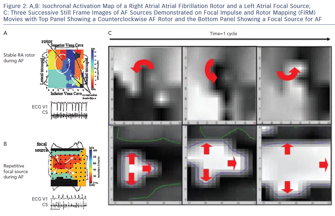
It became clear in early studies that unipolar, bipolar and MAP signals in AF differ dramatically,33 with examples of signals that appeared local in bipolar recordings actually being far-field, i.e. they lay within repolarisation indicated by the MAP.33 This may explain why optical maps of AF, which are based on local action potentials, show rotational drivers of AF in nearly all studies, while traditional electrode based AF activation maps mostly show only disordered activity.8,34,35 These findings, combined with data on conduction velocity and validation of mechanisms by patient-specific targeted ablation, led to Focal Impulse and Rotor Mapping/Modulation (FIRM).27,36,37 An example of a stable right atrial AF driver and a left atrial focal source is shown in Figure 2, where basket catheters are used to identify localised AF drivers. Theoretically, baskets can resolve 1–2 cm diameter reentry circuits in human AF as predicted by Allessie et al., and shown in human optical maps of AF.7,38 Conversely, if electrodes are too closely spaced to map fibrillatory conduction, calculated wave propagation may fall within measurement error. For instance, for atrial conduction velocity in AF patients of 40 cm/sec,39,40 reported errors in assigning onset time in AF (≈5–10 ms) translate to a distance of 2–4 mm (=40×0.005 to 40×0.010). Closer electrode separation than this may attempt to identify circuits within measurement noise, and will have less confidence. A related issue is that closely spaced electrode arrays typically cover small distances simultaneously, which may also miss the 1–2 cm diameter circuits in human AF, noted in optical maps.7,41

Reconciling Differences Between Mapping Studies of AF Rotational Drivers

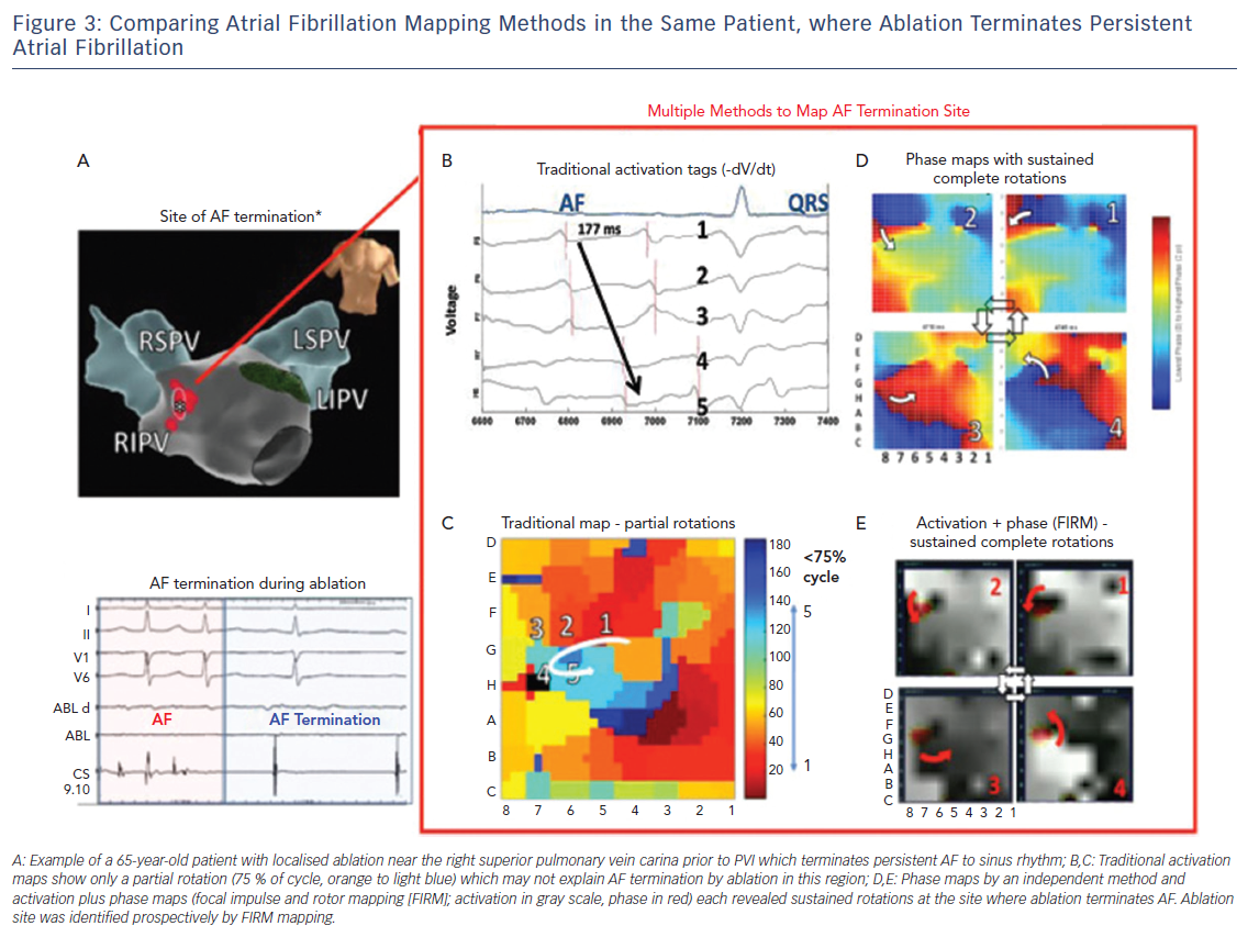
Since long-term ablation success varies dramatically between centres even for a well-defined, anatomic guided technique, such as, due to numbers of lesions, their durability, disease progression and other factors, reconciling differences in AF mapping may be assisted by other study designs.42 On the basis that sites where ablation terminates persistent AF may have mechanistic relevance, we have recently systematically compared maps created by different techniques for analysing the same raw electrographic data in cases of clear termination of persistent AF by ablation, as part of the international COMParison of Algorithms for Rotational Evaluation in AF (COMPARE-AF) study (NCT02997254).43

Figure 3 shows a patient in whom limited ablation before PVI terminated persistent AF to sinus rhythm. In panels C–E, detailed analysis of raw AF electrograms prior to ablation revealed sustained rotational activation by three methods. Of these methods, traditional activation maps of AF electrograms may be confused by spurious deflections, perhaps reflecting far field, and often showed only disorder at AF termination sites where phase and combined phase/activation maps showed rotations.

Long-term Outcomes of AF Driver-Guided Ablation

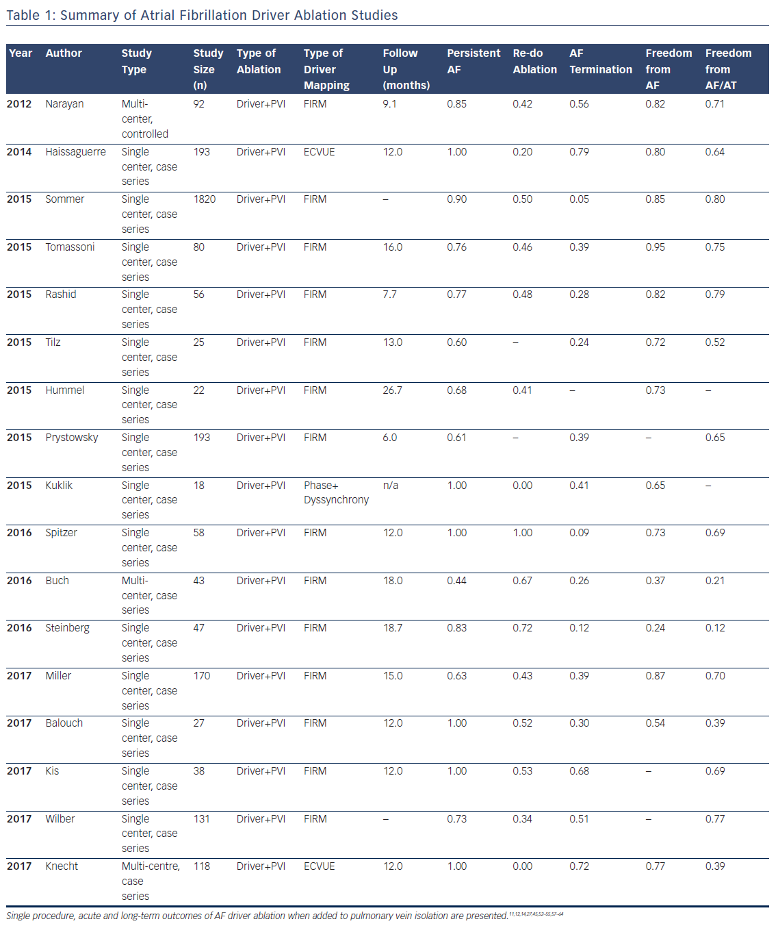
Multiple studies now exist on the outcomes of AF driver ablation (Table 1), with FIRM being the most widely applied technique. As with most new approaches, while initial reports were promising, some recent reports have been disappointing, such as Buch et al. reporting 21 % success and Steinberg et al. 12 % success. These results are surprisingly low since these series included many patients with paroxysmal AF and also performed PVI.44,45

Table 1 summarises studies in a total cohort of 1181 patients undergoing AF driver ablation in addition to PVI. Of these patients, 78 % had non-paroxysmal AF, and the overall single procedure freedom from AF and from AF/all atrial arrhythmias was 76 % and 62 %, respectively. It is noted that reading AF rotational/focal source maps and/or guiding ablation accordingly are novel skills and not learned as part of traditional PVI. Indeed, lower success was seen in studies listed in Table 1 that had fewer cases per operator, and in patients studied early (pre-2013) when no automatic tools were available to interpret maps to assist the operator in this early learning phase. Recent analyses confirm the incremental benefit of adding AF driver ablation to PVI, and there are several multicenter trials ongoing which may provide more clarity on this issue.46

Repetitive focal Source During AF

Article image

Comparing Atrial Fibrillation MApping Methods

Article image

Atrial Fibrillation Driver ABlation Studies

Article image

In most studies, AF drivers arise in diverse locations, overall with 25–40 % near pulmonary veins, 25–40 % elsewhere in the left atrium, and 25–40 % in the right atrium. Body surface mapping (ECVUE) show similar AF driver distributions but in larger regions,11 which may represent greater ‘meander’ in projecting from the heart to the torso.47 In multiple studies, right atrial drivers are mostly in the free wall, posterolateral to the right atrial appendage, and rarely near the superior vena cava or cavotricuspid isthmus. AF drivers are present in higher numbers and more widely distributed in patients with persistent than paroxysmal AF.27

Atrial fibrillation rotational/focal driver sites have been compared to complex fractionated atrial electrogram (CFAE), and AF driver site areas are typically smaller than areas covered by CFAE. However, the concordance between AF drivers and CFAE differs between studies that may reflect differences in identifying CFAE or true differences between methods. FIRM identified sites do not show a specific CFAE grade or voltage fingerprint,27 i.e. CFAE arise at some AF driver sites but are prevalent elsewhere. Conversely, sites identified by body surface mapping could be related to CFAE.48 At the current time, electrogram markers for AF drivers are the subject of intense research.

Practical Approach in Ablation of AF Drivers

Core components required for effective driver-guided ablation include an effective broad-area mapping of both atria, precise identification of drivers to target, and ensuring full elimination of target areas. Each of these components is fertile for technical improvement, and may explain much of the heterogeneity in outcomes between centres.

The FIRM approach to widely map the atria uses multipolar contact basket catheters to analyze many wave-fronts at the same time. In the right atrium, the basket catheter is unsheathed in the superior vena cava (SVC) and slowly retracted into the right atrial body. Slight clockwise or counterclockwise torque is applied for optimal deployment. Particular care is required if a right atrial pacing lead is present, rotating the basket so that splines span the lead rather than displace it, and typically advancing from the inferior vena cava rather than pulling down from the SVC.

In left atrium, the basket is unsheathed in the left superior pulmonary vein and slowly retracted into the atrial body. Slight clockwise or counterclockwise torque may help achieve optimal basket deployment, maximising endocardial contact with the fewest splines over the mitral valve orifice. Alternatively, the basket catheter can be carefully ‘reflected’ off the carina of the left pulmonary veins.

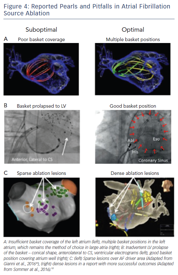
Figure 4 shows examples of optimal and suboptimal basket placement. Various basket sizes and types are available from an increasing number of vendors. We select the most appropriate basket size based on atrial size from intracardiac echocardiography or computed tomography. Figure 4B shows a suboptimal basket positioning that could be identified with fluoroscopic landmarks.49 Importantly, comprehensive atrial coverage may not be possible with a single basket position. Repeat maps after repositioning to sample previously poorly covered regions may help to reveal residual AF drivers.

Successful driver ablation should fully cover the affected driver areas, and contact force sensing catheters may help in this regard. Incomplete coverage may explain lower success rates in some AF driver ablation studies. For instance, Figure 4C shows sparse lesions that may not have eliminated drivers in the long-term, even if AF terminates acutely.50 Incompletely eliminated driver regions may cause recurrent post-ablation AF or atrial tachycardia (AT). Thus, it is critical to ablate the entire affected region, although the science on how much atrium to ablate has yet to be fully established.

Reported Pearls & Pitfalls in AF Source Ablation

Article image

Finally, AF driver-guided ablation does not appear to increase complication rates over traditional ablation alone,51 and is likely not pro-arrhythmic.12,44,52–56

In summary, the endpoint of AF driver ablation is the elimination of sustained rotational or focal activity that lie in conserved spatial locations. This endpoint is achievable in most patients, and may be superior to historical endpoints of AF termination. Lack of AF termination may reflect many mechanisms, including residual AF driver, but clinical results have been promising even in such patients if drivers are eliminated.

Limitations of AF Driver Mapping

While basic and translational science for AF drivers is mature, additional studies are required to reconcile technical differences in mapping for individual patient types. Clinically, large multicentre randomised trials are currently not available, but are ongoing. A learning curve exists in optimum basket placement, interpretation of AF maps, and ablation guidance based on maps. Each of these elements is a subject both for technical innovation and for clinical studies.

Conclusion

Evidence continues to mount that human AF is maintained by rotational and focal sources, and that targeting these areas may improve outcomes over PVI alone. We have outlined the scientific rationale for AF driver ablation, and practical strategies for ablation associated with promising outcomes in multicentre studies. Analysis of less promising studies suggests that suboptimal basket placement, resulting in greater difficulty in reading maps and in targeting ablation, and early learning curve experience may explain at least some of these discrepancies. Future improvements in AF driver ablation may be facilitated by better computational interpretation of AF maps, comparative studies between potentially complementary mapping approaches, and improvements in basket design. Combined mechanistic and imaging studies may enable better and functional classification of AF that may enable better patient tailoring of AF ablation. Ultimately, a better mechanistic and clinical understanding of AF may pave the way for novel drug discovery or regenerative therapies for AF. This is an exciting prospect.

Clinical Perspective

  • AF drivers have been shown in animal and human optical mapping studies, as well as using several new mapping technologies.
  • Studies demonstrate promising outcomes with rotor ablation when added to pulmonary vein isolation.
  • Future improvements to the field would include better automated interpretation of AF maps, comparative studies between mapping approaches that may be complementary, and improved basket designs.

References

  1. Verma A, Jiang CY, Betts TR, et al. Approaches to catheter ablation for persistent atrial fibrillation. N Engl J Med 2015;372:1812–22.
    Crossref | PubMed
  2. Wong KC, Paisey JR, Sopher M, et al. No Benefit OF Complex Fractionated Atrial Electrogram (CFAE) Ablation in Addition to Circumferential Pulmonary Vein Ablation and Linear Ablation: BOCA Study. Circ Arrhythm Electrophysiol 2015;8:1316–24.
    Crossref | PubMed
  3. Calkins H. Demonstrating the value of contact force sensing: more difficult than meets the eye. Circulation 2015;132:901–3.
    Crossref | PubMed
  4. Dukkipati SR, Cuoco F, Kutinsky I, et al. Pulmonary vein isolation using the visually guided laser balloon: A prospective, multicenter, and randomized comparison to standard radiofrequency ablation. J Am Coll Cardiol 2015;66:1350–60.
    Crossref | PubMed
  5. Kuck KH, Brugada J, Furnkranz A, et al. Cryoballoon or radiofrequency ablation for paroxysmal atrial fibrillation. N Engl J Med 2016;374:2235–45.
    Crossref | PubMed
  6. Vogler J, Willems S, Sultan A, et al. Pulmonary vein isolation versus defragmentation: The CHASE-AF clinical trial. J Am Coll Cardiol 2015;66:2743–52.
    Crossref | PubMed
  7. Hansen BJ, Zhao J, Csepe TA, et al. Atrial fibrillation driven by micro-anatomic intramural re-entry revealed by simultaneous sub-epicardial and sub-endocardial optical mapping in explanted human hearts. Eur Heart J 2015;36:2390–401.
    Crossref | PubMed
  8. Pandit SV and Jalife J. Rotors and the dynamics of cardiac fibrillation. Circ Res 2013;112:849–62.
    Crossref | PubMed
  9. Narayan SM, Baykaner T, Clopton P, et al. Ablation of rotor and focal sources reduces late recurrence of atrial fibrillation compared with trigger ablation alone: extended follow-up of the CONFIRM trial (Conventional Ablation for Atrial Fibrillation With or Without Focal Impulse and Rotor Modulation). J Am Coll Cardiol 2014;63:1761–8.
    Crossref | PubMed
  10. Miller MA, Gangireddy SR, Doshi SK, et al. Multicenter study on acute and long-term safety and efficacy of percutaneous left atrial appendage closure using an epicardial suture snaring device. Heart Rhythm 2014;11;1853–9.
    Crossref | PubMed
  11. Haissaguerre M, Hocini M, Denis A, et al. Driver domains in persistent atrial fibrillation. Circulation 2014;130:530–8.
    Crossref | PubMed
  12. Rashid H and Sweeney A. Approaches for focal impulse and rotor mapping in complex patients: A US private practice perspective. J Innovations Card Rhythm Management 2015;6:2193–98.
    Crossref
  13. Tomassoni G, Duggal S, Muir M, et al. Long-term follow-up of FIRM-guided ablation of atrial fibrillation: A single center experience. The Journal of Innovations in Cardiac Rhythm Management. 2015;6:2145–51
  14. Miller JM, Kalra V, Mithilesh DK, et al. Clinical benefit of ablating localized sources for human atrial fibrillation: The Indiana University FIRM Registry. J Am Coll Cardiol 2017;69:1247–56.
    Crossref | PubMed
  15. Haissaguerre M, Jais P, Shah DC, et al. Spontaneous initiation of atrial fibrillation by ectopic beats originating in the pulmonary veins. N Engl J Med 1998;339:659–6.
    Crossref | PubMed
  16. Narayan SM, Bode F, Karasik PL, et al. Alternans of atrial action potentials as a precursor of AF. Circulation 2002;106:1968–73.
    PubMed
  17. Patterson E, Po SS, Scherlag BJ, et al. Triggered firing in pulmonary veins initiated by in vitro autonomic nerve stimulation. Heart Rhythm 2005;2:624–31.
    Crossref | PubMed
  18. Katritsis DG, Giazitzoglou E, Zografos T, et al. Rapid pulmonary vein isolation combined with autonomic ganglia modification: a randomized study. Heart Rhythm 2011;8:672–8.
    Crossref | PubMed
  19. Engelman ZJ, Trew ML and Smaill BH. Structural heterogeneity alone is a sufficient substrate for dynamic instability and altered restitution. Circ Arrhythm Electrophysiol 2010;3:195–203.
    Crossref | PubMed
  20. Narayan SM, Kazi D, Krummen DE, et al. Repolarization and activation restitution near human pulmonary veins and atrial fibrillation initiation: a mechanism for the initiation of atrial fibrillation by premature beats. J Am Coll Cardiol 2008;52:1222–30.
    Crossref | PubMed
  21. Narayan SM, Franz MR, Clopton P, et al. Repolarization alternans reveals vulnerability to human atrial fibrillation. Circulation 2011;123:2922–30.
    Crossref| PubMed
  22. Gonzales MJ, Vincent KP, Rappel W-J, et al. Structural contributions to fibrillatory rotors in a patient-derived computational model of the atria. Europace 2014;16:iv3-iv10.
    Crossref | PubMed
  23. Marrouche NF, Wilber D, Hindricks G, et al. Association of atrial tissue fibrosis identified by delayed enhancement MRI and atrial fibrillation catheter ablation: the DECAAF study. JAMA 2014;311:498–506.
    Crossref | PubMed
  24. Moe GK, Rheinboldt W, Abildskov J. A computer model of atrial fibrillation. Am Heart J 1964;67:200–220.
    PubMed
  25. January CT, Wann LS, Alpert JS, et al. 2014 AHA/ACC/HRS Guideline for the Management of Patients With Atrial Fibrillation: A Report of the American College of Cardiology/American Heart Association Task Force on Practice Guidelines and the Heart Rhythm Society. J Am Coll Cardiol 2014;64: e1–76.
    Crossref | PubMed
  26. Herweg B, Kowalski M, Steinberg JS. Termination of persistent atrial fibrillation resistant to cardioversion by a single radiofrequency application. Pacing Clin Electrophysiol 2003;26:1420–3.
    PubMed
  27. Narayan SM, Krummen DE, Shivkumar K, et al. Treatment of atrial fibrillation by the ablation of localized sources: CONFIRM trial. J Am Coll Cardiol 2012;60:628–36.
    Crossref | PubMed
  28. Shivkumar K, Ellenbogen KA, Hummel JD, et al. Acute termination of human atrial fibrillation by identification and catheter ablation of localized rotors and sources: first multicenter experience of focal impulse and rotor modulation (FIRM) ablation. J Cardiovasc Electrophysiol 2012;23:1277–85.
    Crossref | PubMed
  29. Haissaguerre M, Hocini M, Sanders P, et al. Catheter ablation of long-lasting persistent atrial fibrillation: clinical outcome and mechanisms of subsequent arrhythmias. J Cardiovasc Electrophysiol 2005;16:1138–47.
    Crossref | PubMed
  30. Gray RA, Pertsov AM, Jalife J. Spatial and temporal organization during cardiac fibrillation. Nature 1998;392:75–8.
    Crossref | PubMed
  31. Davidenko JM, Pertsov AV, Salomonsz R, et al. Stationary and drifting spiral waves of excitation in isolated cardiac muscle. Nature 1992;355:349–51.
    Crossref | PubMed
  32. Zlochiver S, Yamazaki M, Kalifa J, et al. Rotor meandering contributes to irregularity in electrograms during atrial fibrillation. Heart Rhythm 2008;5:846–54.
    Crossref | PubMed
  33. Narayan SM, Wright M, Derval N, et al. Classifying fractionated electrograms in human atrial fibrillation using monophasic action potentials and activation mapping: evidence for localized drivers, rate acceleration and non-local signal etiologies. Heart Rhythm 2011;8:244–53.
    Crossref | PubMed
  34. Allessie MA, de Groot NM, Houben RP, et al. Electropathological substrate of long-standing persistent atrial fibrillation in patients with structural heart disease: longitudinal dissociation. Circ Arrhythm Electrophysiol 2010;3:606–15.
    Crossref | PubMed
  35. Lee S, Sahadevan J, Khrestian CM, et al. Simultaneous biatrial high-density epicardial mapping of persistent and long-standing persistent AF in patients: New insights into the mechanism of Its maintenance. Circulation 2015;132:2108–17.
    Crossref | PubMed
  36. Narayan SM, Krummen DE, Rappel W-J. Clinical mapping approach to identify rotors and focal beats in human atrial fibrillation. J Cardiovasc Electrophysiol 2012;23:447–54.
    Crossref | PubMed
  37. Narayan SM, Krummen DE, Enyeart MW, et al. Computational mapping approach identifies stable and long-lived electrical rotors and focal sources in human atrial fibrillation. PLos One 2012;7:e46034.
    Crossref | PubMed
  38. Allessie MA, Bonke FIM, Schopman FJG. Circus movement in rabbit atrial muscle as a mechanism of tachycardia. III: the “leading circle” concept: a new model of circus movement in cardiac tissue without the involvement of an anatomical obstacle. Circ Res 1977;41:9–18.
    PubMed
  39. Lalani G, Schricker A, Gibson M, et al. Atrial conduction slows immediately before the onset of human atrial fibrillation: a bi-atrial contact mapping study of transitions to atrial fibrillation. J Am Coll Cardiol 2012;59:595–606.
    Crossref | PubMed
  40. Schricker A, Rostamian A, Lalani G, et al. Human atrial fibrillation initiates by organized not disorganized mechanisms. Circ Arrhythm Electrophysiol 2014;7:816–24.
    Crossref | PubMed
  41. Zhao J, Hansen BJ, Csepe TA, et al. Integration of high-resolution optical mapping and 3-dimensional micro-computed tomographic imaging to resolve the structural basis of atrial conduction in the human heart. Circ Arrhythm Electrophysiol 2015;8:1514–7.
    Crossref | PubMed
  42. Ganesan AN, Shipp NJ, Brooks AG, et al. Long-term outcomes of catheter ablation of atrial fibrillation: a systematic review and meta-analysis. J Am Heart Assoc 2013;2:e004549.
    Crossref | PubMed
  43. Alhusseini M, Vidmar D, Meckler GL, et al. Two independent mapping techniques identify rotational activity patterns at sites of local termination during persistent atrial fibrillation. J Cardiovasc Electrophys 2017;28:615–22.
    Crossref | PubMed
  44. Buch E, Share M, Tung R, et al. Long-term clinical outcomes of focal impulse and rotor modulation for treatment of atrial fibrillation: a multi-center experience. Heart Rhythm 2016;13:636–41.
    Crossref | PubMed
  45. Steinberg JS, Shah Y, Bhatt A, et al. Focal impulse and rotor modulation: acute procedural observations and extended clinical follow-up. Heart Rhythm 2016;14:192–7.
    Crossref | PubMed
  46. Ramirez FD, Birnie DH, Nair GM, et al. Efficacy and safety of driver-guided catheter ablation for atrial fibrillation: A systematic review and meta-analysis. J Cardiovasc Electrophysiol 2017.
    Crossref | PubMed
  47. Rodrigo M, Guillem MS, Climent AM, et al. Body surface localization of left and right atrial high frequency rotors in atrial fibrillation patients: a clinical-computational study. Heart Rhythm 2014;11:1584–91.
    Crossref | PubMed
  48. Zahid S, Cochet H, Boyle PM, et al. Patient-derived models link re-entrant driver localization in atrial fibrillation to fibrosis spatial pattern. Cardiovasc Res 2016;110:443–54.
    Crossref | PubMed
  49. Benharash P, Buch E, Frank P, et al. Quantitative analysis of localized sources identified by focal impulse and rotor modulation mapping in atrial fibrillation. Circ Arrhythm Electrophysiol 2015;8:554–61.
    Crossref | PubMed
  50. Gianni C, Di Biase L, Deneke T, et al. Acute and short-term outcomes in persistent and long-standing persistent patients undergoing rotors only ablation (Abstract). Heart Rhythm 2015;12:PO01–58
  51. Krummen DE, Baykaner T, Swarup V, et al. Multicenter procedural safety of FIRM ablation (Abstract). Europace 2016
  52. Tomassoni G, Duggal S, Muir M, et al. Long-term follow-up of FIRM-guided ablation of atrial fibrillation: A single-center experience. J Innovations Card Rhythm Management 2015;6:2145–51.
    Crossref
  53. Sommer P, Kircher S, Rolf S, et al. Successful repeat catheter ablation of recurrent longstanding persistent AF with rotor elimination as the procedural endpoint: A case series. J Cardiovasc Electrophysiol 2016;27:274–80.
    Crossref | PubMed
  54. Spitzer SG, Karolyi L, Rammler C, et al. Treatment of recurrent non-paroxysmal atrial fibrillation using focal impulse and rotor mapping (FIRM)-guided rotor ablation: early recurrence and long-term outcomes. J Cardiovasc Electrophysiol 2017;28:31–38.
    Crossref | PubMed
  55. Tilz RR, Lin T, Rillig A, et al. Focal impulse and rotor modulation for the treatment of atrial fibrillation: locations and one year outcomes of human rotors identified using a 64-electrode basket catheter. J Cardiovasc Electrophysiol 2017;28:367–74.
    Crossref
  56. Miller JM, Das MK, Jain R, et al. Clinical benefit of ablating localized sources for human atrial fibrillation: the Indiana University FIRM Registry. J Am Coll Cardiol 2017;69:1247–56.
    Crossref | PubMed
  57. Hoummse M, Hummel J, Hamam I, et al. Long term outcome of FIRM guided rotor ablation for atrial fibrillation. Heart Rhythm Scientific Sessions 2015
  58. Foreman J, Padanilam BJ, Prystowsky EN. Long term outcomes from Focal Impulse and Rotor Modulation for Atrial Fibrillation: A study of 150 cases (Abstract). Heart Rhythm 2015
  59. Kuklik P, Schaeffer B, Hoffmann B, et al. Catheter ablation of persistent atrial fibrillation guided by electrogram dyssynchrony: acute and long term effect. Heart Rhythm Society Scientific Sessions 2015;PO04–101
  60. Buch E, Share M, Tung R, et al. Long-term clinical outcomes of focal impulse and rotor modulation for treatment of atrial fibrillation: A multicenter experience. Heart Rhythm 2015;13:636–41.
    Crossref | PubMed
  61. Balouch M, Gucuk Ipek E, Chrispin J, et al. Impact of rotor temperospatial stability on acute and one-year atrial fibrillation ablation outcomes. Clin Cardiol 2017;40:383–89.
    Crossref | PubMed
  62. Kis Z, Theuns D, Bhagwandien R, et al. Type and rate of atrial fibrillation termination due to rotational activity ablation combined with pulmonary vein isolation. J Cardiovasc Electrophys 2017;28:862–9.
    Crossref | PubMed
  63. Wilber D. FIRM ablation as an adjunct to pulmonary vein isolation. Western AF Symposium, Park City, UT. 2017
  64. Knecht S, Sohal M, Deisenhofer I, et al. Multicentre evaluation of non-invasive biatrial mapping for persistent atrial fibrillation ablation: the AFACART study. Europace 2017;19:1302–9.
    Crossref | PubMed
  65. Gianni C, Mohanty S, Di Biase L, et al. Acute and early outcomes of FIRM-guided rotors-only ablation in patients with non-paroxysmal atrial fibrillation. Heart Rhythm 2016;13:830–5.
    Crossref | PubMed