Article

The Changing Paradigm in the Treatment of Structural Heart Disease and the Need for the Interventional Imaging Specialist

Register or Login to View PDF Permissions
Permissions× For commercial reprint enquiries please contact Springer Healthcare: ReprintsWarehouse@springernature.com.

For permissions and non-commercial reprint enquiries, please visit Copyright.com to start a request.

For author reprints, please email rob.barclay@radcliffe-group.com.
Average (ratings)
No ratings
Your rating

Abstract

Percutaneous interventions in structural heart diseases are emerging rapidly. The variety of novel percutaneous treatment approaches and the increasing complexity of interventional procedures are associated with new challenges and demands on the imaging specialist. Standard catheterisation laboratory imaging modalities such as fluoroscopy and contrast ventriculography provide inadequate visualisation of the soft tissue or three-dimensional delineation of the heart. Consequently, additional advanced imaging technology is needed to diagnose and precisely identify structural heart diseases, to properly select patients for specific interventions and to support fluoroscopy in guiding procedures. As imaging expertise constitutes a key factor in the decision-making process and in the management of patients with structural heart disease, the sub-speciality of interventional imaging will likely develop out of an increased need for high-quality imaging.

Disclosure:HK, FK, RB and RJS have no conflicts of interest to declare. NCW is a consultant for BioVentrix (USA).

Received:

Accepted:

Correspondence Details:Nina C Wunderlich, Cardiovascular Center Darmstadt, Dieburgerstrasse 31c, 64287 Darmstadt, Germany. E: wunderlich@kardio-darmstadt.de

Copyright Statement:

The copyright in this work belongs to Radcliffe Medical Media. Only articles clearly marked with the CC BY-NC logo are published with the Creative Commons by Attribution Licence. The CC BY-NC option was not available for Radcliffe journals before 1 January 2019. Articles marked ‘Open Access’ but not marked ‘CC BY-NC’ are made freely accessible at the time of publication but are subject to standard copyright law regarding reproduction and distribution. Permission is required for reuse of this content.

Over the past 30 years, several percutaneous transcatheter technologies and devices for interventions in structural heart diseases (SHDs) have been introduced (see Table 1). There are numerous technologies that are in development or are currently being used to treat patients with SHD that use transcatheter techniques. The variety of percutaneous treatment approaches has led to a revolution and evolution in clinical care. The past few years have seen a greater application of novel, catheter-based treatments for SHDs. Many of these non-surgical catheter-based interventional procedures have proven effective, which has consequently resulted in a greater acceptance among physicians and patients alike.

The rapid growth of minimally invasive interventional procedures for treating SHD has been accompanied by new challenges and demands on the imaging specialist. Fluoroscopy is inadequate for visualising cardiac valves, congenital and acquired defects. Soft tissue imaging and three-dimensional (3D) delineation of the heart structures is not feasible with fluoroscopy or standard contrast ventriculography. Consequently, additional imaging technology is needed to diagnose and precisely identify SHD in order to appropriately select patients for specific interventions and to support fluoroscopy in guiding procedures.

Three-dimensional transoesophageal echocardiography (TOE) and multi-slice computed tomography (MSCT) provide precise pre-treatment information on anatomic abnormalities, the exact localisation of the cardiac pathology, its severity and precise anatomic dimensions of the lesion.1,2 Such information facilitates procedural planning on the best ways to access and treat the SHD lesion. Moreover, detailed information is provided regarding the anatomy in relation to neighbouring structures.

New technologies for the treatment of SHD are proliferating; consequently, the number of interventions for SHD is increasing3–5 and the procedures are becoming more complex. This has produced a need for greater expertise in the evaluation and imaging of SHD. Due to these changes in the therapeutic armamentarium, interventional imaging appears to be becoming a requisite sub-speciality.

Imaging Modalities for Structural Heart Disease Procedures

Two-dimensional (2D) TOE (or alternatively 2D intracardiac echocardiography) in conjunction with fluoroscopy and cineangiography has been the standard imaging practice in most catheterisation laboratories; however, these imaging modalities are limited in the visualisation of soft tissue, complex 3D structures and their relationships. The introduction of 3D TOE, cardiovascular magnetic resonance (CMR) imaging and MSCT has overcome some of these limitations and, as a result, these additional modalities are being widely adopted in the process of selecting SHD patients, monitoring and guiding procedures, and assessing procedural results. The development and implementation of advanced cardiac imaging constitutes one of the key factors in the success of SHD interventions.6

Three-dimensional Transoesophageal Echocardiography

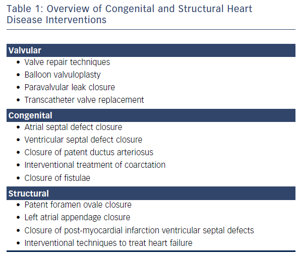
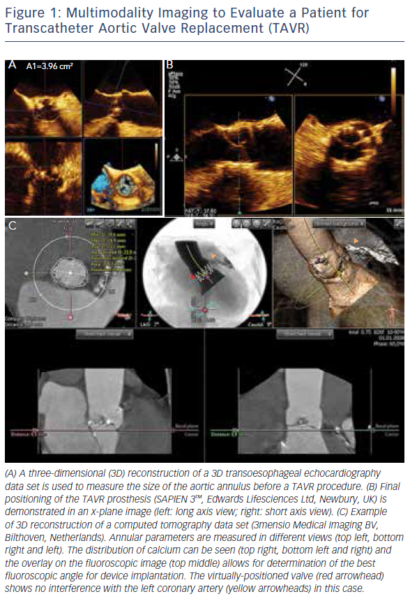
The introduction of 3D TOE has led to a major technological advance in echocardiographic imaging. Realtime 3D TOE imaging provides unique en face views and excellent detail of patients’ 3D anatomy and soft tissue structures. It also allows for the ‘live’ guidance of interventional procedures. Wires, catheters, sheaths, devices and target structures can be seen in one single view and in relation to each other, thus facilitating the guidance of standard and complex SHD interventions. While 3D TOE is gaining acceptance, it is still not available in all centres. Two-dimensional TOE has the advantage of providing higher resolution images, which can be helpful in specific situations where precise measurements are needed. Echo guidance with 2D in conjunction with 3D TOE is crucial and is recommended for many different kinds of mitral intervention,7–10 aortic and mitral paravalvular leak closure,10–13 transcatheter aortic valve replacement (TAVR) (Figure 1A and B),10,14–16 left atrial appendage (LAA) occlusion (Figure 2),10,17 atrial septal defect10,18–20 and patent foramen ovale closure10,21 as well as for percutaneous closure of ventricular septal defects.22

Table 1: Overview of Congenital and Structural Heart Disease Interventions

Article image

Figure 1: Multimodality Imaging to Evaluate a Patient for Transcatheter Aortic Valve Replacement (TAVR)

Article image

Multi-slice Computed Tomography and Cardiovascular Magnetic Resonance Imaging

Although TOE is the most widely used imaging modality, MSCT and CMR are helpful imaging techniques for the pre- and postprocedural assessment of anatomy, pathology and function of cardiac structures, device assessment, and for the detection of complications post-procedure.

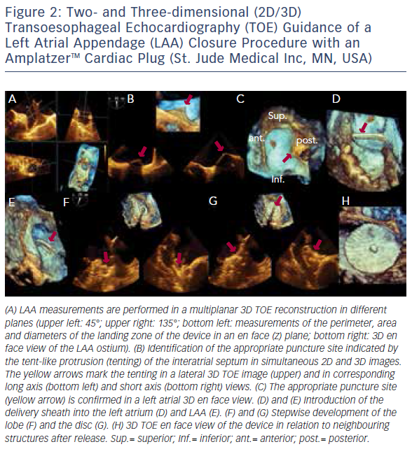
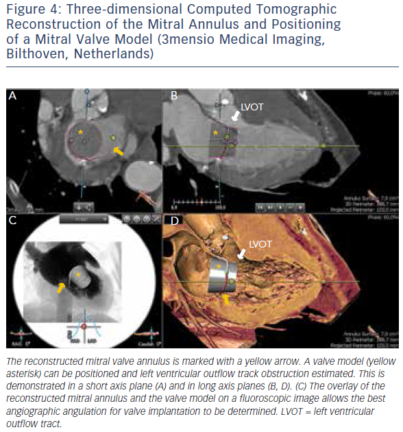
MSCT provides accurate data for sizing devices for TAVR (Figure 1C) and LAA occlusion (Figure 3A–D).2 Before a TAVR procedure, the diameters, areas and perimeters of the aortic annulus, sinuses of Valsalva and ascending aorta can be accurately determined. In addition, the location and distance of the coronary artery ostia from the aortic valve and bypass grafts can reliably be visualised. The amount and distribution of calcification can be identified. MSCT-based analysis of the aortic root and vascular access sites has become routine in the pre-procedural evaluation of patients for TAVR.23–28 In post-procedural assessment, MSCT has been beneficial in detecting complications like reduced aortic-valve leaflet motion in patients with bioprosthetic aortic valves.29 In addition, MSCT is useful for the evaluation of LAA anatomy and measurements regarding device selection, assessment of procedural success and longer-term outcomes.30,31 MSCT is also beneficial in other SHD procedures. It has been used to assess the function and anatomy of the mitral valve complex.32–34 As new mitral valve devices undergo clinical trials, MSCT will likely play a critical role in determining patient eligibility (see Figure 4 for an example of this), especially in the assessment of patients with mitral annular calcification.33,35 Investigators have shown that MSCT can be used to diagnose and differentiate interatrial shunts.36,37 In patients with paravalvular leaks, ECG-gated computed tomography angiography has proven useful in characterising the precise anatomy of the paravalvular leaks, thus facilitating appropriate occluder device selection.38,39

Limitations of MSCT include ionising radiation, a lower temporal resolution than TOE, and the inability to use it during interventional procedures. Nonetheless, due to the proven utility of MSCT in the context of SHD interventions, this imaging modality will likely play a role in the development of future sizing algorithms.

There is limited experience with CMR; however, it is an attractive alternative imaging modality in pre- and post-procedural evaluation of SHD, particularly in patients with renal failure40 and in patients with difficult echocardiographic windows.41 This non-invasive imaging modality allows for detailed visualisation of cardiac anatomy and functional assessment, including quantification of chamber size and volume, left ventricular function (systolic and diastolic), myocardial tissue characterisation and precise wall motion analysis without exposing the patient to ionising radiation.

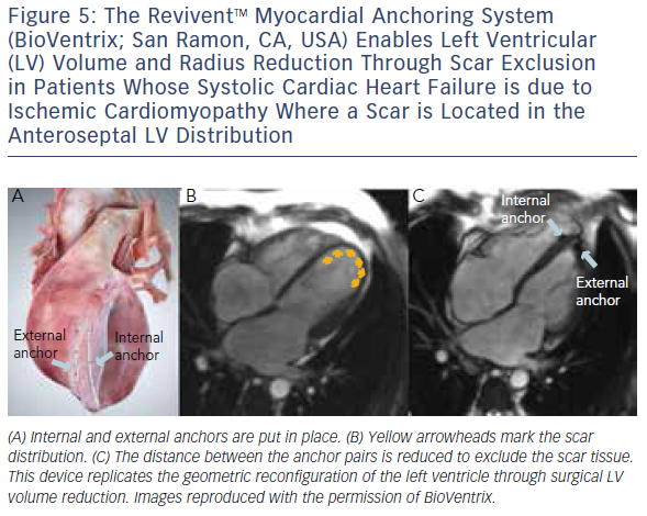
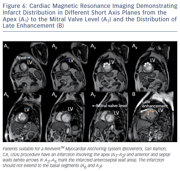
Similar to cardiovascular MSCT, CMR also provides imaging with excellent spatial resolution and can perform 3D multiplanar reconstruction. Figures 5 and 6 demonstrate the usefulness of CMR imaging in patient selection for the ReviventTM Myocardial Anchoring System (BioVentrix, San Ramon, CA, USA). There are, however, limitations to MSCT. These include prolonged examination times, dependence on the ability to perform adequate breath holds, and its limited use in patients with implanted pacemakers or defibrillators and during the device implantation procedure.

Figure 2: Two- and Three-dimensional (2D/3D) Transoesophageal Echocardiography (TOE) Guidance of a Left Atrial Appendage (LAA) Closure Procedure with an AmplatzerTM Cardiac Plug (St. Jude Medical Inc, MN, USA)

Article image

Integrated Multimodality Imaging

More recently, advances in software and hardware development have enabled the integration of various imaging modalities into a single data set, thus resulting in realtime fusion imaging after the separate acquisition of two image data sets. The side-by-side registration of data rendered by different non-invasive imaging modalities such as echocardiography, advanced computed tomography and magnetic resonance imaging technologies and fluoroscopy may overcome some of the limitations of each of the modalities when used as a sole imaging method. Such a multimodal imaging approach may thereby provide increased diagnostic and procedural accuracy by combining anatomical and functional information (see Figure 1C, Figure 3D and E and Figure 4C).42–48

Challenges during Structural Heart Defect Interventions

Currently, interventional transcatheter techniques are being used to treat patients with SHD, including those at high surgical risk. SHD interventions require specifically-designed diagnostic catheters, guiding catheters, guide wires, sheaths and dedicated implantation tools and devices. The visualisation of moving wires, catheters, sheaths and devices within the 3D space of the moving (beating) heart in relation to the target regions constitutes one of the major procedural challenges. In this context, 3D TOE imaging facilitates the manipulation and alignment of devices to the target lesion, thereby increasing the odds of achieving procedural success.

Figure 3: Measurements of the Left Atrial Appendage (LAA) Ostium in a Three-dimensional Computed Tomography Reconstruction and Integration of Structures of Interest into a Fluoroscopic Image by Using the 3mensio Structural Heart Software

Article image

The dynamic and interactive nature of SHD interventions requires a multidisciplinary team with expertise in imaging and intervention. It is crucial that the imaging specialist and the interventionalist communicate with each other at all times during an interventional procedure to ensure optimal and safe deployment of the device. The orientation of echocardiography and fluoroscopy images differs significantly. TOE images are typically obtained through a relatively narrow imaging window through the oesophagus,49 whereas the rotation of the C-arm allows for the acquisition of multiple views of the same structure.35 Thus, the same structure is seen from different perspectives by the echocardiographer and the interventionalist. Identifying the same structure simultaneously on echocardiographic and fluoroscopic images is complicated, therefore the echocardiographer and the interventionalist must make sure that they are communicating effectively to ensure effective manoeuvring of devices to obtain the best possible results.50 As each imaging technique provides unique and supplemental information, the combination of multiple imaging techniques is helpful in precisely defining the anatomy and facilitating device deployment. Thus, the accurate description of anatomy, pathology and function and their effective communication between team members are key factors in a successful procedure. The use of realtime multimodality fusion imaging facilitates this process.42,43

Figure 4: Three-dimensional Computed Tomographic Reconstruction of the Mitral Annulus and Positioning<br />
of a Mitral Valve Model

Article image

The ReviventTM Myocardial Anchoring System

Article image

The Evolution of Interventional Imaging as a New Sub-specialty

Performing SHD interventions is challenging; therefore accurate information on the exact location and anatomy of the target lesions and structures is required. Furthermore, imaging is important for determining the most suitable access for transcatheter procedures. This enables interventionalists to safely and accurately insert and position guide wires, catheters and dedicated devices during structural heart interventions.

Figure 6: Cardiac Magnetic Resonance Imaging Demonstrating

Article image

Due to the complexity of the visual–spatial relationships, imaging specialists are key members of the interventional heart team. As with all cardiovascular procedures, experience is requisite to develop the skills necessary in the clinical setting. Imagers as well as interventionalists performing SHD interventions therefore require specific training on the use of each unique interventional device, as well as knowledge on the specific implantation requirements of these devices.3 Specific guidelines and recommendations, particularly for TOE monitoring of interventional procedures, have been produced by various cardiac societies.3,10,49,51,52 These documents emphasise the value of interventional imaging.

As there are numerous devices available for the treatment of SHD and more in development, substantial growth in the field is expected. Consequently, interventional imaging as a sub-specialty is likely to become even more important than it currently is and is likely to develop into a well defined and recognised sub-specialty.

Conclusion

SHD intervention is a burgeoning field. The sub-specialty of interventional imaging will likely develop out of an increased need for high-quality imaging. Imaging expertise constitutes a key factor in the decisionmaking process and in the management of patients with SHD in order to offer patients optimal outcomes from transcatheter interventions.

References

  1. Faletra FF, Pedrazzini G, Pasotti E, et al. 3D TEE during catheter-based interventions. JACC Cardiovasc Imaging 2014;7:292–308.
    Crossref | PubMed
  2. Xu B, Gooley R, Seneviratne SK, Nasis A. Clinical utility of multi-detector cardiac computed tomography in structural heart interventions. J Med Imaging Radiat Oncol 2016. epub ahead of press
    Crossref | PubMed
  3. Feldman T, Ruiz CE, Hijazi ZM. The SCAI Structural Heart Disease Council: toward addressing training, credentialing, and guidelines for structural heart disease intervention. Catheter Cardiovasc Interv 2010;76:E87–9.
    Crossref | PubMed
  4. Cubeddu RJ, Inglessis I, Palacios IF. Structural heart disease interventions: an emerging discipline in cardiovascular medicine. J Invasive Cardiol 2009;21:478–82.
    PubMed
  5. Herrmann HC, Baxter S, Ruiz CE, et al; SCAI Council on Structural Heart Disease. Results of the Society of Cardiac Angiography and Interventions survey of physicians and training directors on procedures for structural and valvular heart disease. Catheter Cardiovasc Interv 2010;76:E106–10.
    Crossref | PubMed
  6. Carminati M, Agnifili M, Arcidiacono C, et al. Role of imaging in interventions on structural heart disease. Expert Rev Cardiovasc Ther 2013;11:1659–76.
    Crossref | PubMed
  7. Wunderlich NC, Beigel R, Siegel RJ. Management of mitral stenosis using 2D and 3D echo-Doppler imaging. JACC Cardiovasc Imaging 2013;6:1191–205.
    Crossref | PubMed
  8. Wunderlich NC, Siegel RJ. Peri-interventional echo assessment for the MitraClip procedure. Eur Heart J Cardiovasc Imaging 2013;14:935–49.
    Crossref | PubMed
  9. Swaans MJ, Van den Branden BJ, Van der Heyden JA, et al. Three-dimensional transoesophageal echocardiography in a patient undergoing percutaneous mitral valve repair using the edge-to-edge clip technique. Eur J Echocardiogr 2009;10:982–3.
    Crossref | PubMed
  10. Lang RM, Badano LP, Tsang W, et al; American Society of Echocardiography; European Association of Echocardiography. EAE/ASE recommendations for image acquisition and display using three-dimensional echocardiography. J Am Soc Echocardiogr 2012;25:3–46.
    Crossref | PubMed
  11. Krishnaswamy A, Kapadia SR, Tuzcu EM. Percutaneous paravalvular leak closure-imaging, techniques and outcomes. Circ J 2013;77:19–27.
    Crossref | PubMed
  12. Pibarot P, Hahn RT, Weissman NJ, Monaghan MJ. Assessment of paravalvular regurgitation following TAVR: a proposal of unifying grading scheme. JACC Cardiovasc Imaging 2015;8:340–60.
    Crossref | PubMed
  13. Wei J, Yin WH, Lee YT, et al. Intraoperative three-dimensional transesophageal echocardiography for assessing the defect geometries of mitral prosthetic paravalvular leak during transcatheter closure. J Chin Med Assoc 2015;78:158–63.
    Crossref | PubMed
  14. Hahn RT, Gillam LD, Little SH. Echocardiographic imaging of procedural complications during self-expandable transcatheter aortic valve replacement. JACC Cardiovasc Imaging 2015;8:319–36.
    Crossref | PubMed
  15. Hahn RT, Little SH, Monaghan MJ, et al. Recommendations for comprehensive intraprocedural echocardiographic imaging during TAVR. JACC Cardiovasc Imaging 2015; 8:261–87.
    Crossref | PubMed
  16. Shibayama K, Mihara H, Jilaihawi H, et al. 3D assessment of features associated With transvalvular aortic regurgitation after TAVR: A real-time 3D TOE study. JACC Cardiovasc Imaging 2016;9:114–23.
    Crossref | PubMed
  17. Wunderlich NC, Beigel R, Swaans MJ, et al. Percutaneous interventions for left atrial appendage exclusion: options, assessment, and imaging using 2D and 3D echocardiography. JACC Cardiovasc Imaging 2015;8:472–88.
    Crossref | PubMed
  18. Taniguchi M, Akagi T, Kijima Y, Sano S. Clinical advantage of real-time three-dimensional transesophageal echocardiography for transcatheter closure of multiple atrial septal defects. Int J Cardiovasc Imaging 2013;29:1273–80.
    Crossref | PubMed
  19. Roberson DA, Cui VW. Three-dimensional transesophageal echocardiography of atrial septal defect device closure. Curr Cardiol Rep 2014;16:453.
    Crossref | PubMed
  20. Seo JS, Song JM, Kim YH, et al. Effect of atrial septal defect shape evaluated using three-dimensional transesophageal echocardiography on size measurements for percutaneous closure. J Am Soc Echocardiogr 2012;25:1031–40.
    Crossref | PubMed
  21. Martin-Reyes R, López-Fernández T, Moreno-Yangüela M, et al. Role of real-time three-dimensional transoesophageal echocardiography for guiding transcatheter patent foramen ovale closure. Eur J Echocardiogr 2009;10:148–50.
    Crossref | PubMed
  22. Mercer-Rosa L, Seliem MA, Fedec A, et al. Illustration of the additional value of real-time 3-dimensional echocardiography to conventional transthoracic and transesophageal 2-dimensional echocardiography in imaging muscular ventricular septal defects: does this have any impact on individual patient treatment? J Am Soc Echocardiogr 2006;19:1511–9.
    Crossref | PubMed
  23. Achenbach S, Delgado V, Hausleiter J, et al. SCCT expert consensus document on computed tomography imaging before transcatheter aortic valve implantation (TAVI)/transcatheter aortic valve replacement (TAVR). J Cardiovasc Comput Tomogr 2012;6:366–80.
    Crossref | PubMed
  24. Arnold M, Achenbach S, Pfeiffer I, et al. A method to determine suitable fluoroscopic projections for transcatheter aortic valve implantation by computed tomography. J Cardiovasc Comput Tomogr 2012;6:422–8.
    Crossref | PubMed
  25. Jabbour A, Ismail TF, Moat N, et al. Multimodality imaging in transcatheter aortic valve implantation and post-procedural aortic regurgitation: comparison among cardiovascular magnetic resonance, cardiac computed tomography, and echocardiography. J Am Coll Cardiol 2011;58:2165–73.
    Crossref | PubMed
  26. Jilaihawi H, Kashif M, Fontana G, et al. Cross-sectional computed tomographic assessment improves accuracy of aortic annular sizing for transcatheter aortic valve replacement and reduces the incidence of paravalvular aortic regurgitation. J Am Coll Cardiol 2012;59:1275–86.
    Crossref | PubMed
  27. Schultz C, Rossi A, van Mieghem N, et al. Aortic annulus dimensions and leaflet calcification from contrast MSCT predict the need for balloon post-dilatation after TAVI with the Medtronic CoreValve prosthesis. EuroIntervention 2011;7:564–72.
    Crossref | PubMed
  28. Stortecky S, Heg D, Gloekler S, et al. Accuracy and reproducibility of aortic annulus sizing using a dedicated three-dimensional computed tomography reconstruction tool in patients evaluated for transcatheter aortic valve replacement. EuroIntervention 2014;10:339–46.
    Crossref | PubMed
  29. Makkar RR, Fontana G, Jilaihawi H, et al. Possible subclinical leaflet thrombosis in bioprosthetic aortic valves. N Engl J Med 2015;373:2015–24.
    Crossref | PubMed
  30. Vaitkus PT, Wang DD, Guerrero M, et al. Left atrial appendage closure with Amplatzer septal occluder in patients with atrial fibrillation: CT-based morphologic considerations. J Invasive Cardiol 2015;27:258–62.
    PubMed
  31. Wang Y, Di Biase L, Horton RP, et al. Left atrial appendage studied by computed tomography to help planning for appendage closure device placement. J Cardiovasc Electrophysiol 2010;21:973–82.
    Crossref | PubMed
  32. Delgado V, Tops LF, Schuijf JD, et al. Assessment of mitral valve anatomy and geometry with multislice computed tomography. JACC Cardiovasc Imaging 2009;2:556–65.
    Crossref | PubMed
  33. Blanke P, Dvir D, Cheung A, et al. Mitral annular evaluation with CT in the context of transcatheter mitral valve replacement. JACC Cardiovasc Imaging 2015;8:612–5.
    Crossref | PubMed
  34. Van Mieghem NM, Rodríguez-Olivares R, Ren BC, et al. Computed tomography optimised fluoroscopy guidance for transcatheter mitral therapies. EuroIntervention 2016;11:1428–31.
    Crossref | PubMed
  35. Theriault-Lauzier P, Andalib A, Martucci G, et al. Fluoroscopic anatomy of left-sided heart structures for transcatheter interventions: insight from multislice computed tomography. JACC Cardiovasc Interv 2014;7:947–57.
    Crossref | PubMed
  36. Kosehan D, Akin K, Koktener A, et al. Interatrial shunt: diagnosis of patent foramen ovale and atrial septal defect with 64-row coronary computed tomography angiography. Jpn J Radiol 2011;29:576–82.
    Crossref | PubMed
  37. White HD, Halpern EJ, Savage MP. Imaging of adult atrial septal defects with CT angiography. JACC Cardiovasc Imaging 2013;6:1342–5.
    Crossref | PubMed
  38. Kiefer TL, Vavalle J, Hurwitz LM, et al. Resolution of severe hemolysis and paravalvular aortic regurgitation employing an Amplatzer Vascular Plug 4: the importance of detailed pre-procedural planning using CT angiography. Cardiovasc Interv Ther 2015. epub ahead of print
    PubMed
  39. Chiam PT, Ding ZP, Sin YK, et al. How should I treat a percutaneous transcatheter mitral paravalvular leak closure? EuroIntervention 2010;6:653–9.
    Crossref | PubMed
  40. Renker M, Varga-Szemes A, Schoepf UJ, et al. A non-contrast self-navigated 3-dimensional MR technique for aortic root and vascular access route assessment in the context of transcatheter aortic valve replacement: proof of concept. Eur Radiol 2016;26:951–8.
    Crossref | PubMed
  41. Cavalcante JL, Lalude OO, Schoenhagen P, Lerakis S. Cardiovascular magnetic resonance imaging for structural and valvular heart disease interventions. JACC Cardiovasc Interv 2016;9:399–425.
    Crossref | PubMed
  42. Balzer J, Zeus T, Hellhammer K, et al. Initial clinical experience using the EchoNavigator((R))-system during structural heart disease interventions. World J Cardiol 2015;7:562–70.
    Crossref | PubMed
  43. Corti R, Biaggi P, Gaemperli O, et al. Integrated x-ray and echocardiography imaging for structural heart interventions. EuroIntervention 2013;9:863–9.
    Crossref | PubMed
  44. Garcia JA, Bhakta S, Kay J, et al. On-line multi-slice computed tomography interactive overlay with conventional X-ray: a new and advanced imaging fusion concept. Int J Cardiol 2009;133:e101–5.
    Crossref | PubMed
  45. Krishnaswamy A, Tuzcu EM, Kapadia SR. Integration of MDCT and fluoroscopy using C-arm computed tomography to guide structural cardiac interventions in the cardiac catheterization laboratory. Catheter Cardiovasc Interv 2015;85:139–47.
    Crossref | PubMed
  46. Kliger C, Jelnin V, Sharma S, et al. CT angiographyfluoroscopy fusion imaging for percutaneous transapical access. JACC Cardiovasc Imaging 2014;7:169–77.
    Crossref | PubMed
  47. Clegg SD, Chen SJ, Nijhof N, et al. Integrated 3D echo-X ray to optimize image guidance for structural heart intervention. JACC Cardiovasc Imaging 2015;8:371–4.
    Crossref | PubMed
  48. Kim MS, Bracken J, Nijhof N, et al. Integrated 3D Echo-XRay navigation to predict optimal angiographic deployment projections for TAVR. JACC Cardiovasc Imaging 2014;7:847–8.
    Crossref | PubMed
  49. Hahn RT, Abraham T, Adams MS, et al. Guidelines for performing a comprehensive transesophageal echocardiographic examination: recommendations from the American Society of Echocardiography and the Society of Cardiovascular Anesthesiologists. J Am Soc Echocardiogr 2013;26:921–64.
    Crossref | PubMed
  50. Kutty S, Delaney JW, Latson LA, Danford DA. Can we talk? Reflections on effective communication between imager and interventionalist in congenital heart disease. J Am Soc Echocardiogr 2013;26:813–27.
    Crossref | PubMed
  51. Porter TR, Shillcutt SK, Adams MS, et al. Guidelines for the use of echocardiography as a monitor for therapeutic intervention in adults: a report from the American Society of Echocardiography. J Am Soc Echocardiogr 2015;28:40–56.
    Crossref | PubMed
  52. Baumgartner H, Bonhoeffer P, De Groot NM, et al; Task Force on the Management of Grown-up Congenital Heart Disease of the European Society of Cardiology (ESC); Association for European Paediatric Cardiology (AEPC); ESC Committee for Practice Guidelines (CPG). ESC Guidelines for the management of grown-up congenital heart disease (new version 2010). Eur Heart J 2010;31:2915–57.
    Crossref | PubMed