Article

The Brugada Syndrome – Diagnosis, Clinical Implications and Risk Stratification

Register or Login to View PDF Permissions
Permissions× For commercial reprint enquiries please contact Springer Healthcare: ReprintsWarehouse@springernature.com.

For permissions and non-commercial reprint enquiries, please visit Copyright.com to start a request.

For author reprints, please email rob.barclay@radcliffe-group.com.
Average (ratings)
No ratings
Your rating

Abstract

The Brugada syndrome (BrS) is a hereditary arrhythmic syndrome manifesting as syncope or sudden cardiac death (SCD) in individuals without overt structural heart disease. Currently, its diagnosis is mainly based on the presence of a spontaneous or Na+-channel blocker induced so-called “type 1” Brugada electrocardiographic (ECG) pattern typically seen in leads V1 and V2 recorded from the 4th to 2nd intercostal spaces. Presently the main unresolved clinical problem in the BrS is the identification of patients at high risk of SCD who need implantable cardioverter-defibrillator (ICD). Current guidelines recommend ICD implantation only in patients with spontaneous type 1 ECG pattern and either history of aborted cardiac arrest or documented sustained ventricular tachycardia (class I) or syncope of arrhythmic origin (class IIa) because they are at high risk of recurrent arrhythmias. However, the majority of BrS patients are asymptomatic when diagnosed and have generally low risk (0.5 % annually or lower) and therefore are not indicated for ICD. Most of SCD victims in the BrS have had no symptoms prior to the fatal event and therefore were not protected with an ICD. Currently there are no reliable methods to identify these potential victims of SCD. Although some ECG markers such as QRS fragmentation and infero-lateral early repolarisation have been demonstrated to signify increased arrhythmic risk their value still needs to be confirmed in large prospective studies. Novel risk assessment strategies need to be developed based on computerised quantitative ECG analysis of large digital ECG databases in patients with BrS and their relatives, and combined assessment of the most important factors of ventricular arrhythmogenesis.

Disclosure:The author has no conflicts of interest to declare.

Received:

Accepted:

Correspondence Details:Velislav N Batchvarov, Cardiovascular and Cell Sciences Research Institute, St. George’s University of London, Cranmer Terrace, London SW17 0RE E: vbatchva@sgul.ac.uk

Copyright Statement:

The copyright in this work belongs to Radcliffe Medical Media. Only articles clearly marked with the CC BY-NC logo are published with the Creative Commons by Attribution Licence. The CC BY-NC option was not available for Radcliffe journals before 1 January 2019. Articles marked ‘Open Access’ but not marked ‘CC BY-NC’ are made freely accessible at the time of publication but are subject to standard copyright law regarding reproduction and distribution. Permission is required for reuse of this content.

Introduction
The Brugada syndrome (BrS) is a hereditary arrhythmia syndrome manifesting as recurrent syncope or sudden cardiac death (SCD) due to polymorphic ventricular (VT) or fibrillation (VF) in the absence of overt structural heart disease or myocardial ischaemia.1,2 The prevalence of the syndrome is estimated at around 15 per 10,000 in South East Asia including Japan and around 2 per 10,000 in the Western countries.3,4 The BrS may be responsible for up to 4 % of all sudden cardiac deaths (SCD) and at least 20 % of SCDs in patients with structurally normal hearts.5 It is 8–10 times more prevalent in males than in females6 and males are at considerably higher risk of dying suddenly.7 In South-East Asia, the BrS is the leading cause of non-traumatic death in men younger than 40 years.8 This review will briefly summarise current knowledge about the BrS with emphasis on the methods for diagnosis and risk assessment.

Cellular Mechanisms of the BrS
BrS is considered a heritable autosomal dominant disease9 and more than 390 mutations have been identified in the SCN5A gene encoding the α-subunit of the cardiac INa-channel.10 However, presently SCN5A mutations are found only in 11–37 % of the genotyped patients.7,11 Recent data has suggested that heritability may be related to common genetic variation instead of being strictly monogenic.12 Many patients with the BrS have no family history presumably due to under-diagnosis in the other family members, low penetrance or sporadic disease.13

The cellular basis of the BrS is still not fully understood.14 According to the “repolarisation theory”, reduction of the inward Na+ current leads to unopposed transient outward (Ito) current in some epicardial regions of the right ventricular outflow tract (RVOT), which causes either delayed expression of the action potential (AP) dome and epicardial AP prolongation or loss of the dome and AP shortening. The net effect is a potentially arrhythmogenic magnification of repolarisation dispersion between the RVOT endo- and epicardium, and between different RVOT epicardial regions. The repolarisation theory was initially promoted on the basis of experimental studies15,16,17 and was later supported by clinical data such as “spike and dome” configuration with deep notching of monophasic action potentials (MAP) from the RVOT epicardium but not endocardium,18 paradoxical shortening of the RVOT epicardial activation-recovery intervals (ARI) during augmentation of Brugada-type ST segment elevation,19 steep AP duration restitution (slope >1) in the RVOT20,21,22 (both clinically and experimentally), longer ARI in the RVOT epicardium recorded from the conus branch of the right coronary artery than in the endocardium of patients with BrS and type 1 ECG pattern but not in controls23 and others.

There is also mounting evidence from experimental,22 histopathological,24 computational,25 clinical electrophysiological23,25 and imaging26 studies for the presence of conduction abnormalities in the RVOT and their importance for the genesis of ventricular arrhythmias in BrS22,23 (“depolarisation theory”). Delay of the RVOT activation relative to the rest of the RV has also been proposed as a mechanism of the Brugada type ECG changes on the surface ECG.27 The presence of late potentials and prolonged filtered QRS duration on signal-averaged ECG (SAECG) as well as increased notching and fragmentation of the QRS on the standard ECG are linked to increased arrhythmic risk in BrS.28,29,30,31 The reported cases of patients presenting with both arrhythmogenic right ventricular cardiomyopathy (ARVC) and BrS with SCN5A mutations,32 further attest to the likely role of conduction abnormalities in the BrS. A third hypothesis unifying the above two explains the BrS with abnormal expression of the neural crest cells during the embryological development of the RVOT. This defect in the embryogenesis of the RVOT leads to both abnormally augmented electrical gradients during repolarisation as well as to delayed activation of the RVOT.33

From electrocardiographic point of view, the characteristic elevation of the J point and ST segment of the type 1 Brugada ECG pattern (see below) results from early relative (intracellular) positivity of the unaffected zone (RVOT endocardium according to the “repolarisation theory” or normally activated myocardium outside the RVOT according to the “depolarisation theory”), whereas the negative T wave is an expression of late epicardial relative (intracellular) positivity in the affected RVOT zone due to either prolongation of the epicardial APs or its delayed activation.

Clinical Manifestations of the BrS
The symptoms associated with the BrS are due to re-entry ventricular arrhythmias typically arising in the affected zone of the RV. If they last briefly (seconds) and terminate spontaneously they can be asymptomatic or cause palpitations; longer arrhythmias lead to syncope or nocturnal agonal respiration, or can degenerate into VF and cardiac arrest. The duration of the arrhythmia is unpredictable with currently available methods and every arrhythmic episode can be fatal. Therefore, the assessment of the degree of arrhythmic risk and the need for prophylactic treatment is by far the most important aspect of the management of these patients.

The Electrocardiogram – a Key to the Diagnosis of the Brugada Syndrome
The standard 12-lead ECG (with some additional modifications, as explained below) is crucial for the diagnosis and likely also for determining the prognosis (i.e. the level of arrhythmic risk) in the BrS. The diagnostic hallmark of syndrome is the so-called “coved” or “type 1 Brugada ECG pattern” characterised by J-point elevation with slowly descending or concave ST segment elevation merging into a negative or reaching the isoelectric line symmetric T wave7,34 (see Figure 1A). The type 1 Brugada ECG pattern is observed most frequently in leads V1 and V2, much less frequently in lead V3.35 When type 1 Brugada ECG pattern is observed in lead V3 it is always accompanied by the same ECG pattern in at least one more right precordial lead (unpublished observations).

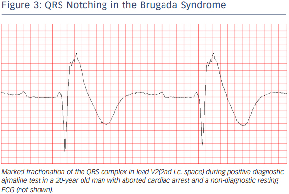
In patients investigated for the BrS, leads V1 and V2 should always be recorded both from the 4th intercostal (i.c.) space as well as from the 3rd and 2nd i.c. space, because the “high” positions increase the sensitivity of leads V1 and V2 for detecting type 1 pattern without loss of specificity36 (see Figure 2). This has been noted already in 1960,37 long before the discovery of the Brugada syndrome. Since the exact anatomic relation between RV and the thoracic cage is individually specific and any of the three i.c. spaces could be closest to the RVOT, it is best to record simultaneously leads V1 and V2 in the 4th, 3rd and 2nd i.c. space. One small study with cardiovascular magnetic resonance imaging found that the maximum RVOT area was most frequently in the 3rd followed by the 4th and 2nd i.c. space.38 In our experience, when type 1 Brugada pattern is observed in the 4th i.c. space it is always also observed in the 3rd or 2nd i.c. space whereas the converse is not the case (unpublished observations). Positioning lead V3 one or two i.c. spaces higher also increases its sensitivity to detect type 1 pattern (unpublished data) (see Figure 2). Bipolar precordial leads between the V2 electrode (positive pole) and V4 or V5 electrodes (negative pole) which can be computed from the standard unipolar leads V2, V4 and V5 seem to be more sensitive and equally specific compared to the unipolar lead V2 for detecting the diagnostic type 1 Brugada ECG pattern.39 Type 1 Brugada ECG pattern sometimes also can be observed in the inferior40,41,42,43 or lateral44,45 leads (the so-called “atypical” BrS).
 

Figure 1: Brugada Type ECG Patterns

Article image


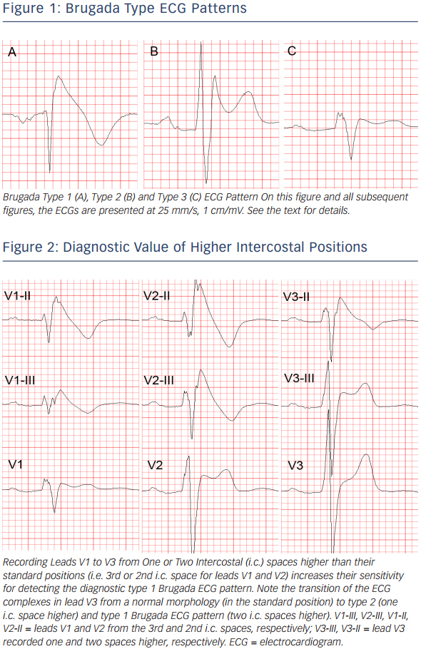
Another ECG pattern of J point and ST segment elevation with a positive T wave in the right precordial leads, the so-called “saddleback” pattern, is considered suspicious but not diagnostic of BrS, unless converted into type 1 pattern following administration of Na+ channel blocking agents. Traditionally the “saddle back” patterns are further divided into “type 2” and “type 3” Brugada ECG pattern depending on the level of J point and ST segment elevation2 (see Figure 1B,C). A recently published expert Consensus Report on the ECG characteristics of the BrS proposed “type 2” and “type 3” patterns to be unified into one “saddle-back” Brugada pattern because, according to the authors’ opinion, the small morphological differences between the two patterns had no diagnostic or prognostic significance.34 In the latest HRS/EHRA/APHRS Expert Consensus Statement on the Diagnosis and Management of Patients with Inherited Primary Arrhythmia Syndromes published in December 2013,7 however, type 2 and type 3 Brugada ECG patterns are still mentioned separately.

Up to 40 % of patients with the BrS present with normal or nondiagnostic resting ECG.46 In these patients, the diagnostic “coved” ECG pattern can be elicited by i.v. administration of sodium channel blocker (ajmaline, procainamide, flecainide).47,48 Currently the BrS is definitely diagnosed when type 1 pattern is observed in at least one of leads V1 and V2 recorded from the 4th, 3rd or 2nd i.c. space either spontaneously or following administration of Na+channel.7 The presence of gene mutations is not considered essential for the diagnosis.2,7

It is important to distinguish between type 2 Brugada ECG pattern and the r’or R’-pattern (incomplete right bundle branch block (iRBBB) pattern) in leads V1 and V2 (especially when recorded from the 3rd or 2nd i.c. space) which can be observed in healthy subjects (frequently in athletes49). It has been reported that a broader angle between the ascending and descending limb of the r’/R’-wave50 or a broader base of the triangle formed by the two limbs of the r’/R’-wave measured at 5 mm from the highest point51,52 can reliably distinguish type 2 Brugada ECG pattern from IRBBB pattern. The classical diagnostic type 1 Brugada ECG pattern needs to be distinguished from similar “Brugadalike” patterns caused by RBBB, septal hypertrophy, arrhythmogenic right ventricular cardiomyopathy (ARVC), pectus excavatum and other conditions, and also from the transient appearance of typical Brugada pattern in the cause of various acute processes such as acute ischaemia, Prinsmetal angina, pulmonary embolism, pericarditis, metabolic disorders, various medications and others (the so-called “Brugada phenocopies”,53 see www.brugadaphenocopy.com). The ECG characteristics of the BrS and the methods to distinguish them from other conditions presenting with similar ECG changes are reviewed in great detail in the above mentioned ECG Consensus Report.34

ECG acquisition with inappropriate high-pass filtering (e.g. non-linear phase high-pass filter of 0.5 Hz instead of the recommended 0.05 Hz)54 can cause considerable ST segment distortion and even mimic type 1 or 2 Brugada pattern.34,55,56

The ECG in BrS characteristically shows considerable dynamic variability; it can be completely normal at one time and demonstrate diagnostic type 1 pattern at another. Vagal influences (slow heart rate, post-prandial state, nighttime) tend to augment the J point and ST segment elevation and the type 1 pattern,57 whereas exercise and catecholamine infusion tend to have the opposite effect (however, in some BrS patients the ST segment elevation might become more prominent during exercise).58 Autonomic influences play important role also in the genesis of malignant arrhythmias because most of the arrhythmic events in BrS occur at night, two long RR intervals often precede episodes of VT/VF,59 whereas catecholamine infusion is used as a first line treatment of such episodes.60 Patients with BrS have increased incidence (10–53 %) of atrial fibrillation (AF).61,62 Additional ECG findings which support the diagnosis of the BrS in asymptomatic patients with spontaneous or induced by Na-channel blockers type 1 pattern include the presence of atrial fibrillation, atrio-ventricular or intraventricular conduction abnormalities (first degree A-V block, fragmented and/or prolonged QRS, abnormal signal-averaged ECG (SAECG), left axis deviation of the QRS complex, prolonged HV interval), ventricular ectopic beats with left bundle branch block (LBBB) morphology and short (<200 ms) ventricular effective refractory period.7

Assessment of the Arrhythmic Risk – the Most Important Clinical Problem in the Brugada Syndrome
The identification of BrS patients with high arrhythmic risk especially among those without previous history of arrhythmia-related symptoms is currently the most important and yet unresolved clinical problem in the BrS. In some aspects, this problem is similar to one of the main (also still not fully resolved) problems of modern cardiology – the identification of patients with ischaemic heart disease (IHD) at high risk of dying suddenly who need prophylactic implantable cardioverter-defibrillator (ICD).

In BrS patients with a previous history of arrhythmic syncope or aborted cardiac arrest the annual event rate of sustained VT or VF is relatively high – between 1.9 %63 and 8.8 %64 and between 7.7 %62 and 13.8 %63, respectively. It is universally accepted that those with aborted cardiac arrest or documented spontaneous sustained VT (with or without syncope) should receive an ICD which is the single therapy with proven efficacy (Class I indication, “…is recommended”).6 There is also evidence that patients with spontaneous type 1 pattern and syncope judged to be of arrhythmic origin (“intermittent risk” group) also are indicated for ICD implantation (currently class IIa indication, “…can be useful.”), whereas asymptomatic patients with spontaneous type 1 pattern are currently considered to represent a “low risk” group (class IIb indication, “…may be considered” depending on the presence or absence of other, not yet fully established risk factors, see below).6 A very recently published Japanese multicentre study confirmed the difference in the level of risk between the latter two patient groups (2.2 % vs 0.5 % during a mean follow-up of 62 months).65

However, the decision to offer an ICD even to a BrS patient with a syncope of presumably arrhythmic origin often is difficult because unlike IHD patients, most of them are relatively young, apparently healthy and without any previous awareness of cardiac problems. Most importantly, it is often very difficult to exclude non-arrhythmic cause of the syncope. In addition, the rate of ICD-related complications (20–30 % annually including inappropriate shocks) is higher than the rate of appropriate activation of the device (2.6–8 % annually).66,67,68 This suggests that novel, better methods of risk-stratification could benefit even some symptomatic BrS patients (i.e. those with the current class IIa indications).6

The majority of BrS patients (64 % in the largest reported series of 1029 BrS patients, the France, Italy, Netherlands, Germany (FINGER) study62 and 63 % in the report of the Brugada syndrome investigators in Japan69) have no symptoms at the time of establishment of the diagnosis. The annual rate of SCD or sustained VT in these patients is low – between 0 %70,71 and 0.872 (0.5 % in the FINGER study62 and the Japanese multicentre study,66 0.4–1 % in several Japanese studies73,74,75) and cannot justify ICD implantation in all of them. On the other hand, the majority of the patients in this heterogeneous group have generally structurally normal hearts and are young or middle aged when diagnosed (median age 45 years in the FINGER study), and therefore the low annual risk can translate into a considerable cumulative arrhythmic risk for the next several decades of their life expectancy. In fact, the majority of victims of SCD in BrS come from this “low risk” (according to current standards) population. One study showed that among patients with the BrS who have died suddenly 68 % had no previous history of arrhythmia-related symptoms and therefore had not been protected by ICD.13 Currently, there are no firmly established reliable methods for the identification of these patients. Similarly, the largest absolute number of patients with IHD who die suddenly also comes from a large patient population considered to have generally low risk (i.e. post-myocardial infarction patients with relatively preserved left ventricular ejection fraction).76

While some studies77,78,79 reported increased occurrence of arrhythmic events in BrS patients with SCN5A mutations these findings have not been confirmed by other studies.80 The genetic analysis is expensive, time-consuming and available only in specialised centres. The role of programmed ventricular stimulation (PVS) during EPS for induction of VT has been an object of controversy and debate since the 1990s. While some early studies supported its value for risk stratification mainly due to its high negative predictive value,81,82 most recent studies failed to confirm its independent predictive value.66,83,84 In the FINGER study,62 inducibility during EPS also did not predict arrhythmic events in multivariate analysis whereas in the multicentre PRELUDE study which tested uniform protocol of PVS during EPS in all 308 patients, the rate arrhythmic events during an average follow-up of three years was not significantly different between the 126 inducible (3.9 %) and the 182 non-inducible patients (4.9 %).85 Currently its role of EPS for risk stratification is accepted only as a Class IIb (“may be considered”) indication.7,86 The method is inherently limited by its invasive character and probably also by the labile nature of the underlying electrophysiologic substrate.87

ECG-derived Parameters for Risk Assessment
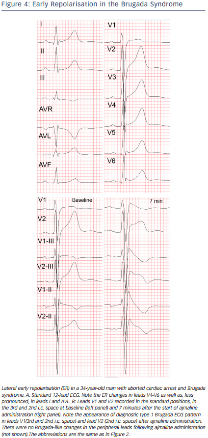
Similarly to other diseases with intraventricular conduction abnormalities such as IHD and cardiomyopathies, the presence of notched or fragmented QRS,31,83 (see Figure 3) has been consistently demonstrated to indicate increased arrhythmic risk independently of other clinical and ECG variables (HR 4.9, 95 % CI 1.6-15.4 in the PRogrammed ELectrical stimUlation preDictive valuE (PERLUDE) study).85 However, currently the presence of notching/fractionation is assessed only visually using arbitrary descriptive criteria (e.g. number of QRS peaks). The presence of late potentials on the signal-averaged ECG (SAECG) is another marker of intraventricular conduction disturbances which also indicates increased arrhythmic risk in BrS30 independently of QRS fractionation. The standard time-domain SAECG, however, cannot detect conduction abnormalities within the QRS complex, has uncertain value in patients with bundle branch block, and uses only a single-lead ECG complex which is derived from the XYZ orthogonal leads and does not contain any regional information.


Figure 3: QRS Notching in the Brugada Syndrome

Article image

Among asymptomatic BrS patients, those with Brugada ECG pattern in the infero-lateral leads in addition to the right precordial leads,88 (increased “spatial Brugada burden”) were recently demonstrated to have several times higher risk of VF compared to those with Brugada type 1 changes only in the right precordial leads.89 On the other hand, the number of right precordial leads displaying type 1 pattern and the degree of J-point or ST-segment elevation do not seem to correlate with the arrhythmic risk.35,36 The “high” (3rd or 2nd i.c. space) positions of leads V1 and V2 are diagnostically more sensitive than their standard positions in the 4th i.c. space90,91,92 but prognostically their value is the same.93

Figure 4: Early Repolarisation in the Brugada Syndrome

Article image

Infero-lateral early repolarisation (ER) (see Figure 4) is not only more common (up to 15 %94) in BrS patients,70,94,95 but also carries up to 4-fold increased risk of spontaneous VF67,70 and even higher risk if the ER has “malignant” morphology,96 i.e. is associated with horizontal/descending (as opposed to rapidly ascending) ST segment after the J point.68,75 The combination of infero-lateral ER and fractionated QRS complex seems to indicate indicate a particularly high arrhythmic risk.97

Currently there are only limited data suggesting that other ECG parameters may indicate increased arrhythmic risk. These parameters include changes in repolarisation dynamics (QT/RR and Tpeak-Tend/RR intervals relations),98 deep negative T wave in lead V1,99< QTc interval more than 460 ms in lead V2 and prolonged Tpeak-Tend interval,100 dynamic alterations in the amplitude of the ST elevation,101 prolonged PR-interval,93 presence of atrial arrhythmias,60,61,102 and augmentation of the ST segment elevation during the early recovery phase of exercise test.103

Summary – Risk Stratification in the Brugada Syndrome – Current Status and Future Diretions
Whereas the diagnosis of the BrS is relatively straightforward with currently available ECG-based methods, the identification of high risk patients who need prophylactic ICD implantation is still an unresolved issue. Currently the only class I indications for ICD implantation in patients diagnosed with the BrS endorsed by the 2013 HRS/EHRA/ APHRS Expert Consensus Statement7 is history of aborted cardiac arrest or documented spontaneous sustained VT, whereas syncope judged to be likely of arrhythmic origin is only a Class IIa indication which mainly reflects the difficulty of excluding a non-cardiac origin of syncope. The guidelines of the Japanese Cardiac Society of 2011 accept practically the same Class I indications whereas for Class IIa indication they require the presence of at least two of the following risk factors: history of syncope, family history of sudden cardiac death and inducible VF during EPS.104 Obviously, these guidelines do not offer solution to the problem of identifying the high risk asymptomatic patients with the BrS. Strict adherence to the HRS/EHRA/APHRS guidelines means that each year in the UK alone, approximately 40 asymptomatic BrS patients are likely to experience their first and potentially lethal arrhythmic event without ICD protection (assuming 0.5 % annual rate in asymptomatic patients [approximately 2/3 of all BrS patients] × estimated 12,600 (2 per 10,000) BrS patients in the UK). Clearly, there is a pressing need to develop novel, easily applicable (e.g. ECG based) risk stratifiers (or combinations thereof) and to confirm prospectively the value of the most promising available ones (e.g. QRS fractionation, infero-lateral ER, possibly others).

Obvious obstacles along the path to this goal are the low rate of arrhythmic events (i.e. end-point events in prospective studies), the small number of patients in the individual centres (since the prevalence of the disease outside South-East Asia is generally low), difficult organisation of big multicentre prospective studies and, possibly, inherent differences between various patient populations (e.g. Western vs Japanese).66 Less appreciated obstacle is the fact that currently ECG research studies (as well as everyday clinical practice) still use mainly 12-lead paper ECGs (or digital ECG images) which are amenable only to visual assessment and simple manual measurement. Computerised mathematical methods for quantitative assessment of QRS and ST-T wave abnormalities have been developed and successfully tested in various cardiac diseases105,106 but they require the availability of digital ECGs (digital files containing the raw ECG signal and not just digital image files). Finally, the development of sustained VT/VF in the BrS is likely a complex event resulting from interaction between the arrhythmic substrate (repolarisation and depolarisation abnormalities) and various triggering and modifying factors (e.g. ventricular ectopic beats, atrial arrhythmias, autonomic modulations such as vagal surge, fever, etc.).14,107,108 Therefore a successful ECG-based risk stratification in BrS should likely involve the combined quantitative assessment of several most important elements of arrhythmogenesis.

References

  1. Brugada P, Brugada J. Right bundle branch block, persistent ST segment elevation and sudden cardiac death: a distinct clinical and electrocardiographic syndrome. J Am Coll Cardiol 1992;20:1391–6.
    Crossref | PubMed
  2. Antzelevitch C, Brugada P, Borggrefe M, et al. Brugada Syndrome. Report of the Second Consensus Conference. Circulation 2005; 111:659–70.
    Crossref | PubMed
  3. Kamakura S. Epidemiology of Brugada syndrome in Japan and rest of the world. Journal of Arrhythmia 2013;29:52–55.
    Crossref
  4. Juang JM, Huang SK. Brugada syndrome—an underrecognized electrical disease 843 in patients with sudden cardiac death. Cardiology 2004;101:157–69.
    Crossref | PubMed
  5. Derval N, Simpson CS, Birnie DH, et al. Prevalence and characteristics of early repolarization in the CASPER registry: cardiac arrest survivors with preserved ejection fraction registry. J Am Coll Cardiol 2011:58:722–8.
    Crossref | PubMed
  6. HRS/EHRA/APHRS Expert Consensus Statement on the Diagnosis and Management of Patients with Inherited Primary Arrhythmia Syndromes. Heart Rhythm 2013;10:1932–63.
    Crossref | PubMed
  7. Kapplinger JD, Tester DJ, Alders M, et al. An international compendium of mutations in the SCN5A-encoded cardiac sodium channel in patients referred for Brugada syndrome genetic testing. Heart Rhythm 2010;7:33–46.
    Crossref | PubMed
  8. Nademanee K, Veerakul G, Nimmannit S, et al. Arrhythmogenic marker for the sudden unexplained death syndrome in Thai men. Circulation 1997;96:2595–600.
    Crossref | PubMed
  9. Horie M, Ohno S. Genetic basis of Brugada syndrome. J Arrhythmia 2013;29:71–6.
    Crossref
  10. The Gene Connection for the Heart. Genetic mutations and inherited arrhythmias. http://www.fsm.it/cardmoc/. Accessed on 24/03/2014.
     
  11. Gehi AK, Duong TD, Metz LD, et al. Risk Stratification of Individuals with the Brugada Electrocardiogram: A Meta- Analysis. J Cardiovasc Electrophysiol 2006;17:577–83.
    Crossref | PubMed
  12. Bezzina CR, Barc J, Mizusawa Y, et al. Common variants at SCN5A-SCN10A and HEY2 are associated with Brugada syndrome, a rare disease with high risk of sudden cardiac death. Nat Genet 2013;45:1044–9.
    Crossref | PubMed
  13. Raju H, Papadakis M, Govindan M, et al. Low Prevalence of Risk Markers in Cases of Sudden Death Due to Brugada Syndrome. Relevance to Risk Stratification in Brugada Syndrome. J Am Coll Cardiol 2011;57:2340–5.
    Crossref | PubMed
  14. Morita M, Zipes DP. Brugada syndrome: Insights of ST elevation, arrhythmogenicity, and risk stratification from experimental observations. Heart Rhythm 2009;6:S34–43.
    Crossref | PubMed
  15. Yan G-X, Antzelevitch C. Cellular Basis for the Brugada Syndrome and Other Mechanisms of Arrhythmogenesis Associated With ST-Segment Elevation. Circulation 1999;100:1660–6.
    Crossref | PubMed
  16. Yan G-X, Shimizu W, Antzelevitch C. Characteristics and Distribution of M Cells in Arterially Perfused Canine Left Ventricular Wedge Preparations. Circulation 1998;98:1921–7.
    Crossref | PubMed
  17. Antzelevitch C. The Brugada syndrome: ionic basis and arrhythmia mechanisms. J Cardiovasc Electrophysiol 2001;12:268–72.
    Crossref | PubMed
  18. Kurita T, Shimizu W, Inagaki M, et al. The Electrophysiologic Mechanism of ST-Segment Elevation in Brugada Syndrome. J Am Coll Cardiol 2002; 40:330–4.
    Crossref | PubMed
  19. Simizu W, Aiba T, Kurita T, Kamakura S. Paradoxic Abbreviation of Repolarization in Epicardium of the Right Ventricular Outflow Tract During Augmentation of Brugada-Type ST Segment Elevation. J Cardiovasc Electrophysiol 2001;12:1418–21.
    Crossref | PubMed
  20. Narayan SM, Kim J, Tate C, Berman BJ. Steep restitution of ventricular action potential duration and conduction slowing in human Brugada syndrome. Heart Rhythm 2007;4:1087–9.
    Crossref | PubMed
  21. Aiba T, Shimizu W, Hidaka I, et al. Cellular Basis for Trigger and Maintenance of Ventricular Fibrillation in the Brugada Syndrome Model High-Resolution Optical Mapping Study. J Am Coll Cardiol 2006;47:2074–85.
    Crossref | PubMed
  22. Lambiase PD, Ahmed AK, Ciaccio EJ, et al. High-Density Substrate Maping in Brugada Syndrome: Combined Role of Conduction and Repolarization Heterogeneities in Arrhythmogenesis. Circulation 2009;120:106–17.
    Crossref | PubMed
  23. Nagase S, Kusano KF, Morita H, et al. Longer Repolarization in the Epicardium at the Right Ventricular Outflow Tract Causes Type 1 Electrocardiogram in Patients With Brugada Syndrome. J Am Coll Cardiol 2008;51:1154–61.
    Crossref | PubMed
  24. Coronel R, Casini S, Koopmann TT, et al. Right Ventricular Fibrosis and Conduction Delay in a Patient With Clinical Signs of Brugada Syndrome. A Combined Electrophysiological, Genetic, Histopathologic, and Computational Study. Circulation 2005;112:2769–77.
    Crossref | PubMed
  25. Nagase S, Kusano KF, Morita H, et al. Epicardial Electrogram of the Right Ventricular Outflow Tract in Patients With the Brugada Syndrome. Using the Epicardial Lead. J Am Coll Cardiol 2002;39:1992–5.
    Crossref | PubMed
  26. Tukkie R, Sogaard P, Vleugels J, et al. Delay in Right Ventricular Activation Contributes to Brugada Syndrome. Circulation 2004;109:1272–7.
    Crossref | PubMed
  27. Meregalli PG, Wilde AAM, Tan HL. Pathophysiological mechanisms of Brugada syndrome: Depolarization disorder, repolarization disorder, or more? Cardiovasc Res 2005;67:367–78.
    Crossref | PubMed
  28. Ikeda T, Sakurada H, Sakabe K, et al. Assessment of Noninvasive Markers in Identifying Patients at Risk in the Brugada Syndrome: Insight Into Risk Stratification. J Am Coll Cardiol 2001;37:1628–34.
    Crossref | PubMed
  29. Furushima H, Chinushi M, Hirono T, et al. Relationship Between Dominant Prolongation of the Filtered QRS Duration in the Right Precordial Leads and Clinical Characteristics in Brugada Syndrome. J Cardiovasc Electrophysiol 2005;16:1311–7.
    Crossref | PubMed
  30. Huang Z, Patel C, MD, et al. Role of signal-averaged electrocardiograms in arrhythmic risk stratification of patients with Brugada syndrome: A prospective study. Heart Rhythm 2009;6:1156–62.
    Crossref | PubMed
  31. Morita H, Kusano KF, Miura D, et al. Fragmented QRS as a Marker of Conduction Abnormality and a Predictor of Prognosis of Brugada Syndrome. Circulation 2008;118:1697-704.
    Crossref | PubMed
  32. Peters S. A second case with arrhythmogenic cardiomyopathy, provocable Brugada ECG and SCN5A mutation. Int J Cardiol 2014;171:e117–8.
    Crossref | PubMed
  33. Elizari MV, Levi R, Acunzo RS, et al. Abnormal expression of cardiac neural crest cells in heart development: A different hypothesis for the etiopathogenesis of Brugada syndrome. Heart Rhythm 2007;4:359–65.
    Crossref | PubMed
  34. Bayés de Luna A, Brugada J, Baranchuk A, Borggrefe M, Breithardt G, Goldwasser D, Lambiase P, et al. Current electrocardiographic criteria for diagnosis of Brugada pattern: a consensus report. J Electrocardiol 2012;45:433–42.
    Crossref | PubMed
  35. Richter S, Sarkozy A, Paparella G, et al. Number of electrocardiogram leads displaying the diagnostic covedtype pattern in Brugada syndrome: a diagnostic consensus criterion to be revised. Eur Heart J 2010;31:1357–64.
    Crossref | PubMed
  36. Govindan M, Batchvarov VN, Raju H, et al. Utility of high and standard right precordial leads during ajmaline testing for the diagnosis of Brugada Syndrome. Heart 2010;96:1904–8.
    Crossref | PubMed
  37. Roesler H. An electrocardiographic study of high take-off of the R(R’)-T segment in right precordial leads. Am J Cardiol 1960;6:920–8.
    Crossref | PubMed
  38. Veltmann CT, Papavassiliu T, Konrad T, Insights into the location of type I ECG in patients with Brugada syndrome: Correlation of ECG and cardiovascular magnetic resonance imaging. Heart Rhythm 2012;9:414–21.
    Crossref | PubMed
  39. Batchvarov VN, Govindan M, Macfarlane PF, et al. Diagnostic Utility of Bipolar Precordial Leads During Ajmaline Testing for Suspected Brugada Syndrome. Heart Rhythm 2010; 7:208–15.
    Crossref | PubMed
  40. Takagi M, Aihara N, Takaki H, et al. Clinical Characteristics of Patients With Spontaneous or Inducible Ventricular Fibrillation Without Apparent Heart Disease Presenting with J Wave and ST Segment Elevation in Inferior Leads. J Cardiovasc Electrophysiol 2000;11:844–8.
    Crossref | PubMed
  41. Chinushi M, Izumi D, Furushima H, et al. Multiple Premature Beats Triggered Ventricular Arrhythmias During Pilsicainide Infusion in a Patient with Inferior ST-Segment Elevation. Pacing Clin Electrophysiol 2006;29:1445–8.
    Crossref | PubMed
  42. Kalla H, Yan G-X, Marinchak R. Ventricular Fibrillation in a Patient with Prominent J (Osborn) Waves and ST Segment Elevation in the Inferior Electrocardiographic Leads: A Brugada Syndrome Variant? J Cardiovasc Electrophysiol 2000;11:95–8.
    Crossref | PubMed
  43. Batchvarov VN, Govindan M, Camm AJ, Behr ER. Brugada- Like Changes in the Peripheral Leads During Diagnostic Ajmaline Test in Patients With Suspected Brugada Syndrome. Pacing Clin Electrophysiol 2009;32:695–703.
    Crossref | PubMed
  44. Van den Berg MP, Wiesfeld ACP. Brugada Syndrome with ST-Segment Elevation in the Lateral Leads. J Cardiovasc Electrophysiol 2006;17:1.
    Crossref | PubMed
  45. Bonakdar H, Haghjoo M, Sadr-Ameli MA. Brugada Syndrome Manifested by the Typical Electrocardiographic Pattern both in the Right Precordial and the High Lateral Leads. Ind Pacing Electrophysiol J 2008;8:137–40.
    Crossref | PubMed
  46. Rivero-Ayerza M, Brugada R, Brugada J, et al. Electrocardiogram of Brugada Syndrome and its Dynamic Patterns. In: Malik M, Camm AJ (editors), Dynamic Electrocardiography, Futura/Blackwell Publishing, NY; 2004;417–24.
    Crossref
  47. Brugada R, Brugada J, Antzelevitch C et al. Sodium channel blockers identify risk for sudden death in patients with ST-segment elevation and right bundle branch block but structurally normal hearts. Circulation 2000;101:510–5.
    Crossref | PubMed
  48. Rolf S, Bruns H-J, Wichter T, et al. The Ajmaline challenge in Brugada syndrome: Diagnostic impact, safety, and recommended protocol. Eur Heart J 2003;24:1104–12.
    Crossref | PubMed
  49. Chung EH, McNeely DE, Gehi AK, et al. Brugada-type patterns are easily observed in high precordial lead ECGs in collegiate athletes. J Electrocardiol 2014;47:1–6.
    Crossref | PubMed
  50. Chevallier S, Forclaz A, Tenkorang J, et al. New Electrocardiographic Criteria for Discriminating Between Brugada Types 2 and 3 Patterns and Incomplete Right Bundle Branch Block. J Am Coll Cardiol 2011;58:2290–8.
    Crossref | PubMed
  51. Serra G, Baranchuk A, Bayes de Luna A, et al. New electrocardiographic criteria to differentiate the Type-2 Brugada pattern from electrocardiogramof healthy athletes with r′-wave in leads V1/V2. Europace 2014;16:1639–45.
    Crossref | PubMed
  52. García-Niebla J, Baranchuck A, Bayés de Luna A. True Brugada pattern or only high V1-V2 electrode placement? J Electrocardiol 2014;47:756–8.
    Crossref | PubMed
  53. Baranchuk A, Nguyen T, Ryu MH, et al. Brugada Phenocopy: New Terminology and Proposed Classification. Ann Noninvas Electrocardiol 2012;17:299–314.
    Crossref | PubMed
  54. Kligfield P, Gettes LS, Bailey JJ, et al. AHA/ACC/HRS Ssinetific Statements. Recommendations for the Standardization and Interpretation of the Electrocardiogram Part I: The Electrocardiogram and Its Technology. J Am Coll Cardiol 2007;49:1109–27.
    Crossref | PubMed
  55. García-Niebla J, Serra-Autonell G, Bayés de Luna A. Brugada syndrome electrocardiographic pattern as a result of improper application of a high pass filter. Am J Cardiol 2012;110:318–20.
    Crossref | PubMed
  56. García-Niebla J, Serra-Autonell G, Fiol M, Bayés de Luna A. Brugada electrocardiographic pattern: Reality or fiction? J Electrocardiol 2014;47:362–3.
    Crossref | PubMed
  57. Mizumaki K, Fujiki A, Nishida K, et al. Bradycardia-dependent ECG changes in Brugada syndrome. Circ J 2006;70:896–901.
    Crossref | PubMed
  58. Amin AS, de Groot EA, Ruijter JM, et al. Exercise-induced ECG changes in Brugada syndrome. Circ Arrhythm Electrophysiol 2009;2:531–9.
    Crossref | PubMed
  59. Matsuo K, Shimizu W, Kurita T, et al. Dynamic changes of 12-lead electrocardiograms in a patient with Brugada syndrome. J Cardiovasc Electrophysiol 1998;9:508–12.
    Crossref | PubMed
  60. Veerakul G, Nademanee K. Treatment of electrical storms in Brugada syndrome. J Arrhythmia 2013;29:117–24.
    Crossref
  61. Morita H, Kusano-Fukushima, Nagase S, et al. Atrial fibrillation and atrial vulnerability in patients with Brugada syndrome. J Am Coll Cardiol 2002;40:1437–44.
    Crossref | PubMed
  62. Kusano KF, Taniyama MK, Nakamura K, et al. Atrial fibrillation in patients with Brugada syndrome relationships of gene mutation, electrophysiology, and clinical backgrounds. J Am Coll Cardiol 2008;51:1169–75.
    Crossref | PubMed
  63. Probst V, Veltmann C, Eckardt L, et al. Long-Term Prognosis of Patients Diagnosed With Brugada Syndrome: Results from the FINGER Brugada syndrome Registry. Circulation 2010;121:635–43.
    Crossref | PubMed
  64. Brugada J, Brugada R, Antzelevitch C, et al. Long-term followup of individuals with the electrocardiographic pattern of right bundle-branch block and ST-segment elevation in precordial leads V1 to V3. Circulation 2002;105:73–8.
    Crossref | PubMed
  65. Takagi M, Sekiguchi Y, Yokoyama Y, et al. for the Japan Idiopathic Ventricular Fibrillation Study (J-IVFS )Investigators. Long-term prognos is inpatients with Brugada syndrome based on Class II indication for implantable cardioverterdefibrillator in the HRS/EHRA/APHRS Expert Consensus Statement: Multicenter study in Japan. Heart Rhythm 2014;11:1716–20.
    Crossref
  66. Sacher F, Probst V, Iesaka Y, et al. Outcome after implantation of a cardioverter-defibrillator in patients with Brugada syndrome: a multicenter study. Circulation 2006;114:2317–24.
    Crossref | PubMed
  67. Sarkozy A, Boussy T, Kourgiannides G, et al. Long-term follow-up of primary prophylactic implantable cardioverterdefibrillator therapy in Brugada syndrome. Eur Heart J 2007;28:334–44.
    Crossref | PubMed
  68. Rosso R, Glick A, Glikson M, et al. Outcome after implantation of cardioverter defibrillator [corrected] in patients with Brugada syndrome: a multicenter Israeli study (ISRABRU). Isr Med Assoc J 2008;10:435–9.
    PubMed
  69. Kamakura S, Ohe T, Nakazawa K, et al. Long-term prognosis of probands with Brugada-pattern ST-elevation in leads V1– V3. Circulat Arrhythmia Electro-Physiol 2009;2:495–503.
    Crossref | PubMed
  70. Letsas KP, Weber R, Efremidis M, et al. Long-term prognosis of asymptomatic individuals with spontaneous or druginduced type 1 electrocardiographic phenotype of Brugada syndrome. J Electrocardiol 2011;44:346–9.
    Crossref | PubMed
  71. Champagne J, Philippon F, Gilbert M, et al. The Brugada syndrome in Canada: a unique French-Canadian experience. Can J Cardiol 2007;23(Suppl B):71B.
    Crossref | PubMed
  72. Eckardt L, Probst V, Smits JP, et al. Long-term prognosis of individuals with right precordial ST-segment-elevation Brugada syndrome. Circulation 2005;111:257.
    Crossref | PubMed
  73. Atarashi H, Ogawa S. Idiopathic Ventricular Fibrillation Investigators. New ECG criteria for high-risk Brugada syndrome. Circ J 2003;67:8.
    Crossref | PubMed
  74. Takagi M, Aonuma K, Sekiguchi Y, et al, The Japan Idiopathic Ventricular Fibrillation Study. The prognostic value of early repolarization (J wave) and ST-segment morphology after J wave in Brugada syndrome: Multicenter study in Japan (J-IVFS) Investigators. Heart Rhythm 2013;10:533–9.
    Crossref | PubMed
  75. Yokokawa M, Noda T, Okamura H, et al. Comparison of longterm follow-up of electrocardiographic features in Brugada syndrome between the SCN5A-positive probands and the SCN5A-negative probands. Am J Cardiol 2007;100:649.
    Crossref | PubMed
  76. Huikuri HV, Castellanos A, Myerburg RJ. Sudden Death Due to Cardiac Arrhythmias. N Engl J Med 2001;345:1473–82.
    Crossref | PubMed
  77. Sommariva E, Pappone C, Martinelli Boneschi F, et al. Genetics can contribute to the prognosis of Brugada syndrome: a pilot model for risk stratification. Eur J Human Genetics 2013;21:911–7.
    Crossref | PubMed
  78. Nishii N,Ogawa M,Morita H, et al.SCN5A mutation is associated with early and frequent recurrence of ventricular fibrillation in patients with Brugada syndrome. Circ J 2010;74:2572–8.
    Crossref | PubMed
  79. Meregalli PG, Tan HL, Probst V, et al. Type of SCN5A mutation determines clinical severity and degree of conduction slowing in loss-of-function sodium channelopathies. Heart Rhythm 2009;6:341–8.
    Crossref | PubMed
  80. Priori SG, Napolitano C, Gasparini M, et al. Natural history of Brugada syndrome: insights for risk stratification and management. Circulation 2002;105:1342–7.
    Crossref | PubMed
  81. Brugada J, Brugada R, Brugada P. Electrophysiologic testing predicts events in Brugada syndrome Patients. Heart Rhythm 2011; 8:1595–7.
    Crossref | PubMed
  82. Sarkozy A, Paparella G, Boussy T, et al. The usefulness of the consensus clinical diagnostic criteria in Brugada syndrome. Int J Cardiol 2013;167:2700–4.
    Crossref | PubMed
  83. Gasparini M, Priori SG, Mantica M, et al. Provocative tests in the Brugada syndrome: do we have the right tools? Circulation 2000;102(suppl2):677.
     
  84. Eckardt L, Kirchhof P, Schulze-Bahr E, et al. Electrophysiologic investigation in Brugada syndrome. Yield of programmed ventricular stimulation at two ventricular sites with up to three premature beats. European Heart Journal 2002;23:1394–401.
    Crossref | PubMed
  85. Priori SG, Gasparini M, Napolitano C, et al. Risk stratification in Brugada syndrome results of the PRELUDE (Programmed Electrical stimUlation preDictive valuE) registry. J Am Coll Cardiol 2012;59:37–45.
    Crossref | PubMed
  86. Zipes DP, Camm AJ, Borggrefe M, et al. ACC/AHA/ESC 2006 guidelines for management of patients with ventricular arrhythmias and the prevention of sudden cardiac death: a report of the American College of Cardiology/American Heart Association Task Force and the European Society of Cardiology Committee for Practice Guidelines. Circulation 2006;114:e385–484.
    Crossref | PubMed
  87. Wichter T, Matheja P, Eckardt L, et al. Cardiac Autonomic Dysfunction in Brugada Syndrome. Circulation 2002;105:702–6.
    Crossref | PubMed
  88. Rollin A, Sacher F, Gourraud JB, et al. Prevalence, characteristics, and prognosis role of type 1 ST elevation in the peripheral ECG leads in patients with Brugada syndrome. Heart Rhythm 2013;10:1012–8.
    Crossref | PubMed
  89. Viskin S, Adler A, Rosso R. Brugada burden in Brugadasyndrome: The way to go in risk stratification? Heart Rhythm 2013;7:1019–20.
    Crossref | PubMed
  90. Shimizu W, Matsuo K, Takagi M, Tanabe Y, Aiba T, Taguchi A, Suyama K, Kurita T, Aihara N, Kamakura S: Body surface distribution and response to drugs of ST segment elevation in Brugada syndrome: clinical implication of eighty-sevenlead body surface potential mapping and its application to twelve-lead electrocardiograms. J Cardiovasc Electrophysiol 2000;11:396–404.
    Crossref | PubMed
  91. Sangwatanaroj S, Prechawat S, Sunsaneewitayakul B, Sitthisook S, et al: New electrocardiographic leads and the procainamide test for the detection of the Brugada sign in sudden unexplained death syndrome survivors and their relatives. Eur Heart J 2001;22:2290–6.
    Crossref | PubMed
  92. Teijeiro R, Garro HA, Acunzo RS, et al. Recording of High V1-V3 Precordial Leads May Be Essential to the Diagnosis of Brugada Syndrome During the Ajmaline Test. J Cardiovasc Pharmacol and Therapeutics 2006;11:153–5.
    Crossref | PubMed
  93. Miyamoto K,Yokokawa, M,Tanaka K, et al. Diagnostic and prognostic value of a type 1 Brugada electrocardiogram at higher (third or second) V1 to V2 recording in men with Brugada syndrome. Am J Cardiol 2007;99:53–7.
    Crossref | PubMed
  94. Sarkozy A, Chierchia GB, Paparella G, et al. Inferior and lateral electrocardio-graphic repolarization abnormalities in Brugada syndrome. Circulat Arrhythmia Electrophysiol 2009;2:154–61.
    Crossref | PubMed
  95. Bastiaenen R, Raju H, Sharma S, et al. Characterization of early repolarisation during ajmaline provocation and exercise tolerance testing. Heart Rhythm 2013;10:247–54.
    Crossref | PubMed
  96. Rosso R, MD, Glikson E, Belhassen B, et al. Distinguishing “benign” from “malignant early repolarization”: The value of the ST-segment morphology. Heart Rhythm 2012;9:225–9.
    Crossref | PubMed
  97. Tokioka K, Kusano KF, Morita H, et al. Electrocardiographic parameters and fatal arrhythmic events in patients with Brugada syndrome: combination of depolarization and repolarization abnormalities. J Am Coll Cardiol 2014;63:2131–8.
    Crossref | PubMed
  98. Sangawa M, Morita H, Nakatsu T, et al. Abnormal transmural repolarization process in patients with Brugada syndrome. Heart Rhythm 2009;6:1163–9.
    Crossref | PubMed
  99. Miyamoto A, Hayashi H, Makiyama T, et al. Risk determinants in individuals with a spontaneous type 1 Brugada ECG. Circ J 2011;75:844–51.
    Crossref | PubMed
  100. Castro Hevia J, Antzelevitch C, Tornes Barzaga F, et al. Tpeak–Tend and Tpeak–Tend dispersion as risk factors for ventricular tachycardia/ventricular fibrillation in patients with the Brugada syndrome. J Am Coll Cardiol 2006;47:1828–34.
    Crossref | PubMed
  101. Take Y, Morita H, Wu J, et al. Spontaneous electrocardiogram alterations predict ventricular fibrillation in Brugada syndrome. HeartRhythm 2011;8:1014–21.
    Crossref | PubMed
  102. Bordachar P, Reuter S, Garrigue S, et al. Incidence, clinical implications and prognosis of atrial arrhythmias in Brugada syndrome. Eur Heart J 2004;25:879–84.
    Crossref | PubMed
  103. Makimoto H, Nakagawa E,Takak I H, et al. Augmented ST-segment elevation during recovery from exercise predicts cardiac events in patients with Brugada syndrome. J Am Coll Cardiol 2010;56:1576–84.
    Crossref | PubMed
  104. Shimizu A. Indication of ICD in Brugada syndrome. J Arrhythmia 2013;29:110–6.
    Crossref | PubMed
  105. Okin PM, Devereux RB, Fabsitz RR, et al. Principal Component Analysis of the T Wave and Prediction of Cardiovascular Mortality in American Indians. The Strong Heart Study. Circulation 2002;105:714–9.
    Crossref | PubMed
  106. Addison PS. Wavelet transforms and the ECG: a review. Physiol Meas 2005; 26:R155–99.
    Crossref | PubMed
  107. Cellular basis for trigger and maintenance of ventricular fibrillation in the Brugada syndrome model: high-resolution optical mapping study. J Am Coll Cardiol 2006;47:2074–85.
    Crossref | PubMed
  108. Morita H, Zipes DP, Lopshire J, et al. T wave alternans in an invitro canine tissue model of Brugada syndrome. Am J Physiol Heart Circ Physiol 2006;291:H421–8.
    Crossref | PubMed